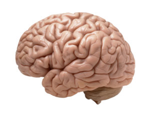
 Another article was published this month raising the issue of whether Alzheimer's disease is caused by a microbe - which can explain why all the medicines and experimental drugs aimed at treating the "tangles" or amyloid plaques in the brain are not working as a treatment (because that's the wrong approach). The microbe theory of Alzheimer's disease has been around for decades, but only recently is it starting to be taken seriously. Some of the microbes found in patients with Alzheimer's disease (from analyses of both normal brains and Alzheimer patient brains after death): fungi, Borrelia burgdorferi (Lyme disease), herpes simplex virus Type 1 (HSV1), and Chlamydia pneumoniae.
The general hypotheses seem to be that Alzheimer’s disease is caused by infection, but it isn't linked to any one pathogenic microbe. Instead, the evidence seems to support that "following infection, certain pathogens gain access to brain, where immune responses result in the accumulation of amyloid-β, leading to plaque formation". So the microbes act as "triggers" for Alzheimer's disease - the microbes get into the brain, and immune responses somehow eventually result in the amyloid plaques and Alzheimer's disease. From The Scientist:
Do Microbes Trigger Alzheimer’s Disease?
In late 2011, Drexel University dermatology professor Herbert Allen was astounded to read a new research paper documenting the presence of long, corkscrew-shape bacteria called spirochetes in postmortem brains of patients with Alzheimer’s disease. Combing data from published reports, the International Alzheimer Research Center’s Judith Miklossy and colleagues had found evidence of spirochetes in 451 of 495 Alzheimer’s brains. In 25 percent of cases, researchers had identified the spirochete as Borrelia burgdorferi, a causative agent of Lyme disease. Control brains did not contain the spirochetes.
Allen had recently proposed a novel role for biofilms—colonies of bacteria that adhere to surfaces and are largely resistant to immune attack or antibiotics—in eczema.... Allen knew of recent work showing that Lyme spirochetes form biofilms, which led him to wonder if biofilms might also play a role in Alzheimer’s disease. When Allen stained for biofilms in brains from deceased Alzheimer’s patients, he found them in the same hippocampal locations as amyloid plaques. Toll-like receptor 2 (TLR2), a key player in innate immunity, was also present in the same region of the Alzheimer’s brains but not in the controls. He hypothesizes that TLR2 is activated by the presence of bacteria, but is locked out by the biofilm and damages the surrounding tissue instead.
Spirochetes, common members of the oral microbiome, belong to a small set of microbes that cross the blood-brain barrier when they’re circulating in the blood, as they are during active Lyme infections or after oral surgery. However, the bacteria are so slow to divide that it can take decades to grow a biofilm. This time line is consistent with Alzheimer’s being a disease of old age, Allen reasons, and is corroborated by syphilis cases in which the neuroinvasive effects of spirochetes might appear as long as 50 years after primary infection.
Allen’s work contributes to the revival of a long-standing hypothesis concerning the development of Alzheimer’s. For 30 years, a handful of researchers have been pursuing the idea that pathogenic microbes may serve as triggers for the disease’s neuropathology..... In light of continued failures to develop effective drugs, some researchers, such as Harvard neurobiologist Rudolph Tanzi, think it’s high time that more effort and funding go into alternative theories of the disease. “Any hypothesis about Alzheimer’s disease must include amyloid plaques, tangles, inflammation—and, I believe, infection.”
Herpes simplex virus type 1 (HSV1) can acutely infect the brain and cause a rare but very serious encephalitis. In the late 1980s, University of Manchester molecular virologist Ruth Itzhaki noticed that the areas of the brain affected in HSV1 patients were the same as those damaged in patients with Alzheimer’s disease. Knowing that herpes can lie latent in the body for long periods of time, she began to wonder if there was a causal connection between the infection and the neurodegenerative disorder.
Around the same time, neuropathologist Miklossy, then at the University of Lausanne in Switzerland, was detailing the brain damage caused by spirochetes—both in neurosyphilis and neuroborrelia, a syndrome caused by Lyme bacteria. She happened upon a head trauma case with evidence of bacterial invasion and plaque formation, and turned her attention to Alzheimer’s. She isolated spirochetes from brain tissue in 14 Alzheimer’s patients but detected none in 13 age-matched controls. In addition, monoclonal antibodies that target the amyloid precursor protein (APP)—which, when cleaved, forms amyloid-β—cross-reacted with the spirochete species found, suggesting the bacteria might be the source of the protein.
Meanwhile, in the U.S., a third line of evidence linking Alzheimer’s to microbial infection began to emerge. While serving on a fraud investigation committee, Alan Hudson, a microbiologist then at MCP-Hahnemann School of Medicine in Philadelphia, met Brian Balin.... Soon, Balin began to send Hudson Alzheimer’s brain tissue to test for intracellular bacteria in the Chlamydia genus. Some samples tested positive for C. pneumoniae: specifically, the bacteria resided in microglia and astrocytes in regions of the brain associated with Alzheimer’s neuropathology, such as the hippocampus and other limbic system areas. Hudson had a second technician repeat the tests before he called Balin to unblind the samples. The negatives were from control brains; the positives all had advanced Alzheimer’s disease. "We were floored,” Hudson says.
Thus, as early as the 1990s, three laboratories in different countries, each studying different organisms, had each implicated human pathogens in the etiology of Alzheimer’s disease. But the suggestion that Alzheimer’s might have some microbial infection component was still well outside of the theoretical mainstream. Last year, Itzhaki, Miklossy, Hudson, and Balin, along with 29 other scientists, published a review in the Journal of Alzheimer’s Disease to lay out the evidence implicating a causal role for microbes in the disease.
The microbe theorists freely admit that their proposed microbial triggers are not the only cause of Alzheimer’s disease. In Itzhaki’s case, some 40 percent of cases are not explained by HSV1 infection. Of course, the idea that Alzheimer’s might be linked to infection isn’t limited to any one pathogen; the hypothesis is simply that, following infection, certain pathogens gain access to brain, where immune responses result in the accumulation of amyloid-β, leading to plaque formation.
 Something surprising: People with multiple sclerosis don't develop Alzheimer's disease - even if it runs in the family. New research suggests that multiple sclerosis may protect a person from Alzheimer's disease.