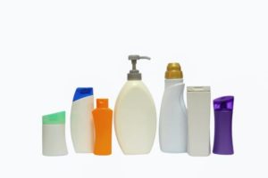
 Flame retardants. All around us, and in us. So, so hard to avoid because they're in electronic goods, in upholstered furniture, polyurethane foam, carpet pads, some textiles, the foam in baby items, house dust, building insulation, and on and on. And unfortunately, while a number of toxic flame retardants have been phased out, it appears that the new replacements may be just as bad and are more easily inhaled (the small particles go down the air tract and into the lung tissue).
What to do? Wash hands before eating. Try to use a vacuum cleaner with a HEPA filter. Try to avoid products that say they contain "flame retardants". Only buy upholstered furniture with tags that say they are flame retardant free. From Environmental Health News:
As Washington state decides on stronger toxics law, residents are breathing flame retardants
A new generation of chemicals added to furniture, building insulation and baby products like car seats to slow the spread of flames are escaping into air at higher levels than previously thought, according to a new study out of Washington state. The findings come as Washington lawmakers decide on bolstering flame retardant bans. The state was one of the first to ban an earlier generation of retardants, known as PBDEs.
The new research found flame retardant chemicals used to replace polybrominated diphenyl ethers (PBDEs) also escape, are ubiquitous in indoor air and suggest inhalation is a major route of exposure for people. The compounds, called chlorinated organophosphate flame retardants, found in the study have been linked to cancer and reproductive problems, and some can alter hormones essential for development. “We’ve been underestimating what total exposure is,” said Erika Schreder, staff scientist at the Washington Toxics Coalition and lead author of the study published this month in the scientific journal Chemosphere.
Researchers gave 10 people from Washington state an air sampler that simulates breathing to wear during a normal day: office work, commuting, hanging out at home. They tested for a suite of the new generation of chlorinated flame retardants and found all 10 were breathing some amount of them throughout the day. Exposure to one of the most prevalent compounds was up to 30 times greater than ingesting the chemicals via dust. The distinction is important: dust exposure occurs largely through the mouth, previously thought to be the major exposure route for banned PBDEs.
Chlorinated flame retardants are used mostly in polyurethane foam, often in building insulation and everyday products such as furniture, children’s car seats and baby strollers. The compounds are substitutes for PBDEs, which were widely used as flame retardants until scientists reported they were building up in people and wildlife and various bans took hold.
While chlorinated flame retardants have been around for decades, Salamova said scientists have recently started to understand them as, at first, it was thought they weren’t harmful or able to accumulate in people and wildlife. However there is evidence the replacement are following the same path as PBDEs: chlorinated flame retardants have been found in household dust, children’s products, drinking water, and mother-toddlers pairs. Two chlorinated flame retardants have been flagged by the state of California as carcinogens, and animal research suggests they may hamper brain development as well.
From Medical Xpress: Prenatal exposure to flame retardants linked to poorer behavioral function in children
New research from the University of Cincinnati (UC) College of Medicine suggests that prenatal exposure to flame retardants and perfluoroalkyl substances (PFASs) commonly found in the environment may have a lasting effect on a child's cognitive and behavioral development, known as executive function...."We examined the relationship between prenatal exposure to PBDEs and PFASs and executive function in children at 5 and 8 years of age," said Ann Vuong, DrPH, a postdoctoral fellow at the University of Cincinnati in the Department of Environmental Health. "The findings suggest that maternal serum concentrations of PBDEs and perfluorooctane sulfonate (PFOS), one of the most commonly found PFASs in human blood, may be associated with poorer executive functioning in school-age children."
From Science Daily: Exposure to common flame retardants may contribute to attention problems in children
Prenatal exposure to some flame retardants that have been widely-used in consumer products is associated with attention problems in young children. A new study is the first to show the effects of prenatal exposure to polybrominated diphenyl ethers on children's development at ages 3, 4, and 7 years. Children with the highest exposure to certain PBDEs had approximately twice the number of maternally-reported attention problems compared to the other children in the study. PBDEs are found in textiles, plastics, wiring, and furniture containing polyurethane foam to reduce flammability.
Very nice and thorough report about flame retardants written in 2013 by the highly regarded center EHHI (Environment and Human Health, Inc.): FLAME RETARDANTS THE CASE FOR POLICY CHANGE
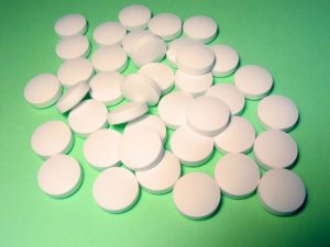
 Once again, research shows that "BPA-free" plastic does not mean it is safer than BPA plastic. Both BPA and BPS (the usual replacement for BPA) leach estrogenic chemicals into the foods and beverages, which means negative health effects when ingested. Both BPA and BPS mimic the effects of estrogen, as well as the actions of thyroid hormone. Yes, this study was done on zebrafish, but think of them as "the canaries in the mine" - if it affects them, it could affect humans also, especially developing fetuses and young children.
 I posted about this amazing research while it was still ongoing (
 This confirms what researchers such as
  Could the bacteria described in this research be another probiotic or beneficial bacteria (besides Lactobacillus sakei) that helps protect against sinusitis? New research found that the harmless bacteria Corynebacterium accolens is "overrepresented" in children free of Streptococcus pneumoniae (pneumococcus) - which commonly colonizes in children's noses (and that can live harmlessly as part of a healthy microbiome), but it is also an important infectious agent. Streptococcus pneumoniae is a major cause of pneumonia, septicemia, meningitis, otitis media (ear infections), and sinusitis in children and adults worldwide.
 More evidence that traditional toys and books are superior to electronic toys in both verbal parent-child interactions and non-verbal interactions for young children. Parent-child verbal interactions are so important because they teach young children language, lay the groundwork for literacy skills, teach role-playing, teach emotional and social skills such as turn-taking and accepting others' leads. In other words, put down the electronic gadgets and go spend time talking and interacting with your young child with old style traditional toys and books. From Science Daily:
 The results of this study lead me to say DUH...of course spending time with children and being responsive to them, talking and interacting a lot with them, being affectionate and loving with them is best. So in this holiday season, don't just give gifts and toys to your children and think you can leave them to their own devices or with others, but spend time with them, talk a lot to them, play games with them, interact, and do things with them. Put down your own electronic devices (cell phone, laptop, tablet, etc) and go spend time with your child. From Science Daily:
 A large study found that using antidepressants during the second or third trimester of pregnancy increases the risk that the child will have autism by 87%, especially if the mother takes selective serotonin reuptake inhibitors (SSRIs). A drawback was that the study looked at associations rather than actual cause (which would have meant randomly assigning women to either treatment or no treatment - which is unethical). From Medical Xpress:
 Just a few years ago the type of pesticide (organophosphate, for example chlorpyrifos) looked at in this study was commonly used everywhere - in schools, homes, agriculture. It was easy to buy in stores (e.g., Raid spray), and was considered "safer" than older pesticides. Over time problem after problem has been found with them - with the latest being decreased lung function in children exposed to organophosphates early in life.