 Another study showing that higher physical activity (from a variety of activities) is "related to larger gray matter volume in the elderly, regardless of cognitive status", specifically in gray matter areas of the brain responsible for memory, learning, and cognition. In other words, higher levels of physical activity reduce brain atrophy that occurs with aging and improves cognitive function in elderly individuals. There is also discussion of higher activity levels improving cerebral (brain) blood flow. Bottom line: get off your butt and move more for better brain health. From Medical Xpress:
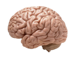
Burning more calories linked with greater gray matter volume, reduced Alzheimer's risk
Whether they jog, swim, garden or dance, physically active older persons have larger gray matter volume in key brain areas responsible for memory and cognition, according to a new study by researchers at the University of Pittsburgh School of Medicine and UCLA.The findings, published today in the Journal of Alzheimer's Disease, showed also that people who had Alzheimer's disease or mild cognitive impairment experienced less gray matter volume reduction over time if their exercise-associated calorie burn was high.
A growing number of studies indicate physical activity can help protect the brain from cognitive decline, said investigator James T. Becker, Ph.D., professor of psychiatry, Pitt School of Medicine..... "Our study is one of the largest to examine the relationship between physical activity and cognitive decline, and the results strongly support the notion that staying active maintains brain health."
Led by Cyrus Raji, M.D., Ph.D., formerly a student at Pitt School of Medicine and now a senior radiology resident at UCLA, the team examined data obtained over five years from nearly 876 people 65 or older participating in the multicenter Cardiovascular Health Study. All participants had brain scans and periodic cognitive assessments. They also were surveyed about how frequently they engaged in physical activities, such as walking, tennis, dancing and golfing, to assess their calorie expenditure or energy output per week.
Using mathematical modeling, the researchers found that the individuals who burned the most calories had larger gray matter volumes in the frontal, temporal and parietal lobes of the brain, areas that are associated with memory, learning and performing complex cognitive tasks. In a subset of more than 300 participants at the Pitt site, those with the highest energy expenditure had larger gray matter volumes in key areas on initial brain scans and were half as likely to have developed Alzheimer's disease five years later.
"Gray matter houses all of the neurons in your brain, so its volume can reflect neuronal health," Dr. Raji explained. "We also noted that these volumes increased if people became more active over five years leading up to their brain MRI."
 Not good news. More than half of Americans’ calories come from “ultra-processed foods,” according to
  Yes, the chemicals in personal care products and cosmetics you use absolutely get into your body, have effects, and can be measured in the urine. Of especially big concern are the endocrine (hormone) disrupting chemicals such as phthalates, parabens, triclosan, and oxybenzone (BP-3). This study shows that even taking a 3 day break from these chemicals lowers their levels in your body. The researchers found that : "The adolescent girls in this study experienced an average within girl decline of 27-45% in urinary concentrations of certain phthalates, certain parabens, triclosan, and oxybenzone after three days of abstaining from conventional personal care products and using replacement products with labels indicating they did not contain these chemicals."
 Two recent studies link low vitamin D levels with more aggressive cancers: aggressive prostate cancer in men and more aggressive breast cancers (in mice and women). Researchers generally advise people to take 1000 to 2000 international units per day of vitamin D3 to maintain normal blood levels of of more than 30 nanograms/milliliter. The best source of vitamin D is sunlight, which is why vitamin D is frequently called the sunshine vitamin.
 For years stores and manufacturers have promoted the advantages of Scotchgard and Teflon nonstick coatings for pots and pans, as stain-proofing for upholstered furniture and rugs, as a water repellent for clothing, for consumer goods such as dental floss, and for grease-proof food wrappers and containers. And yes, people have been convinced - with most cookware sold today being of the nonstick type, and the popularity of sofas and rugs coated with non-stain coatings. But once again, chemicals come with a price and health effects, and unfortunately these particular chemicals are found in all of us in varying levels.
 Flame retardants. All around us, and in us. So, so hard to avoid because they're in electronic goods, in upholstered furniture, polyurethane foam, carpet pads, some textiles, the foam in baby items, house dust, building insulation, and on and on. And unfortunately, while a number of toxic flame retardants have been phased out, it appears that the new replacements may be just as bad and are more easily inhaled (the small particles go down the air tract and into the lung tissue).
_lores.jpg) Borrelia burgdorferi Credit: CDC
Borrelia burgdorferi Credit: CDC