Skip to content

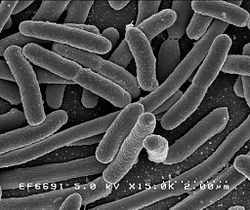
Here is an amazing short video for those interested in seeing how bacteria mutate and grow when exposed to antibiotics - and evolving to become superbugs. Researchers filmed an experiment that created bacteria a thousand times more drug-resistant than their ancestors. In the time-lapse video, a white bacterial colony (E.coli bacteria) creeps across an enormous black petri dish plated with vertical bands of successively higher doses of antibacterial drugs (antibiotics).

How they did it: The researchers imaged the E. coli bacteria every 10 minutes for 10 days as the microbes expanded across the plate. You can see that the bacteria paused briefly at the boundaries of increasingly stronger antibiotic concentrations until a mutant bacteria struck out into the stronger antibiotic territory. By challenging the bacteria with differing doses of antibiotic, the team demonstrated that E. coli evolve higher resistance more quickly if they first encounter an intermediate, rather than a high, concentration of antibiotic. It's a beautiful, yet horrifying video. NOTE: the bacteria grows on agar, which is a thick, clear substance that comes from seaweed and is used for growing bacteria in scientific research. From Harvard Medical School, on YOUTUBE:

From NPR:  WATCH: Bacteria Invade Antibiotics And Transform Into Superbugs

In the time-lapse video, a white bacterial colony creeps across an enormous black petri dish plated with vertical bands of successively higher doses of antibiotic. The colony pauses when it hits the first band of antibiotic, creating a stark border between the white colony and the black petri dish. Then the bacteria start to edge their way into the toxic soup. More dots appear and they start growing, racing to the next, stronger band of antibiotic. The bacteria are evolving. After almost two weeks of real time have passed, they've become resistant to the strongest completely taken over the kitchen-table-sized petri dish.

We know dangerous bacteria are getting stronger all the time and that it's our fault because of our excessive and indiscriminate use of antibiotics. Each year, 23,000 people in the U.S. die as a result of superbug infections. But we typically don't get to see superbugs created.... Their video and report were published Thursday in the journal Science. 

This article by Dr. Thomas E. Finucane lays out nicely a paradigm shift in how to view uncomplicated urinary tract infections (UTIs) - as a case of dysbiosis (microbial community out of whack), and that antibiotics to kill bacteria are generally not needed or helpful. (He doesn't mention it, but the next step in his argument should be that probiotic or beneficial bacteria or other microbes may improve the microbial community and symptoms.) A main point of the article is that we now know the urinary tract is not sterile - instead diverse microbiota live there (the microbial community is the microbiome) including bacteria and viruses (the virome), and that these stable microbial communities are generally beneficial. Standard cultures do not pick up all the microbes living in the urinary tract.

He points out that: UTI symptoms are usually self-limited, of brief duration and only slightly shortened by antibiotic treatment; that cystitis rarely progresses to pyelonephritis (which does need antibiotic treatment); and that randomized trials show no reduction in the risk of progression to pyelonephritis with antibiotic treatment. He stresses the "generally benign (other than symptoms) nature of “symptomatic UTI” is suggested by the billions of persons around the world and over the years who have suffered “UTI” without access to antibiotics and have recovered fully". And that "urinary tract dysbiosis" may be a better description of what a woman is experiencing.

However, I would like to add that to a person experiencing an UTI, the pain does not at all feel "benign". So look at the posts on UTIs and treatments and perhaps try something like D- mannose  or cranberry supplements, or both. From The American Journal of Medicine:

“Urinary tract infection” and the microbiome

The current paradigm for managing uncomplicated “urinary tract infection” (“UTI”) is deeply flawed. “UTI” is ambiguously defined and, coupled with a belief that “bacteria are not normal inhabitants of the urinary tract, the diagnosis often leads to unnecessary, harmful antibiotic treatment. Although bacteriuria identified by standard clinical cultures (which we will call standard bacteriuria) is central to most definitions, more sensitive diagnostic tests now demonstrate that “urine is not sterile2 and that standard bacteriuria represents a fraction of the diverse microbiota hosted by the urinary tract. Knowledge of this complex, generally beneficial microbiome deeply undermines the current paradigm, which relies on the findings of standard culture. By acknowledging this microbiome a successor paradigm will generate new questions about relationships among host, microbiome and antibiotic use and will almost surely show additional serious harms from antibiotic overtreatment.

This discussion concerns medically stable, non-pregnant adults with normal urinary tract structure and function. The role of antibiotics in patients with abnormalities of anatomy or physiology, such as spinal cord injury, urinary obstruction, or catheters, will require careful investigation. New insight into pyelonephritis and bacteremic bacteriuria is likely to develop.

The ambiguous definition of “UTI” seems to promote antibiotic overuse. In one common usage, “urinary tract infection is defined as microbial infiltration of the normally sterile urinary tract.” With this definition, asymptomatic bacteriuria is a “UTI” and is often treated, even in patient groups where strong evidence shows lack of benefit.4 A second common definition, “significant bacteriuria in a patient with symptoms or signs attributable to the urinary tract and no alternate source” seems more restrictive but does not define what symptoms or signs may be attributed to the urinary tract. This ambiguity creates opportunities for overtreatment....Antibiotic treatment of “UTI” often follows even though no data have shown these changes respond to treatment.

Canonically, “all symptomatic UTI should be treated” but actual benefit is limited. Hooton emphasizes that in acute uncomplicated cystitis “the primary goal of treatment is to ameliorate symptoms.” Foxman summarizes that symptoms are usually self-limited, of brief duration and only slightly shortened by antibiotic treatment; that cystitis rarely progresses to pyelonephritis; and that randomized trials show no reduction in the risk of progression to pyelonephritis with antibiotic treatment.7 The generally benign (other than symptoms) nature of “symptomatic UTI” is suggested by the billions of persons around the world and over the eons who have suffered “UTI” without access to antibiotics and have recovered fully.

With its various meanings, convenient diagnosis, long tradition, suggestive link to treatment and uncritical acceptance by clinicians, patients, families and insurers, “UTI” remains heavily embedded in practice, “one of the most common bacterial infections worldwide”. The paradigm provides tidy management for a patient with “UTI” who expects antibiotics. Further, the current paradigm does account for several findings. Standard bacteriuria is associated with pyuria, fever and dysuria, for example, and these often improve with treatment, as do a wide variety of findings seemingly unconnected with the urinary tract. Antibiotic treatment improves outcomes for asymptomatic pregnant women who have standard bacteriuria. Pyelonephritis and bacteremic bacteriuria probably arise in the urinary tract and do require antibiotic treatment.

To diagnose “UTI” and determine antibiotic sensitivity based on results of standard cultures, however, is to rely on familiar, accessible data and to ignore the dozens of bacterial speciesas well as intracellular bacterial colonies and urinary virome known to reside in the urinary tract. Current discussions of symptomatic or asymptomatic bacteriuria or sterile urine are similarly problematic. To attribute delirium to standard bacteriuria seems unjustifiable, knowing that most or all people with or without delirium have bacteriuria. The current paradigm is defensible only if all pathogenic organisms are identified with standard cultures and all organisms more difficult to identify can be safely ignored.

We propose instead that urinary symptoms, bacteremia, pyelonephritis, and other recognizable disturbances of the urinary tract are the dysbiotic tip of a much larger iceberg of complex host-microbe interactions that are occurring out of sight of standard cultures. As expected in the era of the microbiome, stable bacterial communities are generally beneficial. For example, compared with the instillation of sterile saline, “bladder colonization with (the nonpathogenic) E. coli HU2117 safely reduces the risk of symptomatic urinary tract infection in patients with spinal cord injury”.8 Of 699 young women with asymptomatic bacteriuria, half of whom were randomized to receive no antibiotic treatment, “treatment was associated with a higher rate of symptomatic UTI… (thus) asymptomatic bacteriuria … may play a protective role in preventing symptomatic recurrence” during 12-month follow-up.9

Costello and colleagues outline a broader paradigm shift in the general approach to infection; “transitioning clinical practice from the Body-as-Battleground to the Human-as-Habitat perspective will require rethinking how one manages the human body.10 To help in this transition, mindful language will be important. We suggest that authors use “UTI” only within quotation marks and that clinicians use the bimanual “air quotes” gesture in discussions. This small, repetitive annotation is intended to disrupt the term’s complacent usage and encourage rethinking of how one manages bacteriuria. The term “urinary tract dysbiosis” may be useful for otherwise well patients with urinary tract symptoms.

“UTI” is an ill-defined, glibly overdiagnosed and overtreated “infection”. Current management ignores modern science. The associated antibiotic overuse causes serious harm to patient safety and to public health. Instead of the current-paradigm question, “Does this patient have a UTI?” the successor-paradigm question will be, “Does evidence show that antibiotic treatment is likely to benefit this patient?” Shifting the paradigm is an urgent matter.

A recent study of microbiomes (microbial communities) of patients admitted to intensive care units (ICU) found that they had rapid loss of normal, “health promoting” bacteria", which resulted in the "overgrowth of disease-promoting pathogenic bacteria (dysbiosis), which, in turn, makes patients susceptible to hospital-acquired infections, sepsis, and organ failure". In other words, serious illnesses disrupt human microbial communities, as do treatments, medicines, antibiotics, and lack of proper nutrition in intensive care units. Interestingly, they observed "large depletions of organisms previously thought to confer anti-inflammatory benefits, such as Faecalibacterium". Faecalibacterium prausznitzii has been discussed in other posts as an incredibly important beneficial bacteria for health, a keystone species in the gut (here and here).

The researchers, who took skin, oral, and fecal samples at two time points, expressed surprise over how rapidly the microbial communities changed, and suggested that possible treatments for the micobial communities being out-of-whack (dysbiosis) are "probiotics or with targeted, multimicrobe synthetic “stool pills” that restore a healthy microbiome in the ICU setting to improve patient outcomes." In other words, "restoration of a healthy gut microbiome may be important for improving outcomes in critically ill patients".  Of course.... From Science Daily:

ICU patients lose helpful gut bacteria within days of hospital admission

The microbiome of patients admitted to the intensive care unit (ICU) at a hospital differs dramatically from that of healthy patients, according to a new study published in mSphere. Researchers analyzing microbial taxa in ICU patients' guts, mouth and skin reported finding dysbiosis, or a bacterial imbalance, that worsened during a patient's stay in the hospital. Compared to healthy people, ICU patients had depleted populations of commensal, health-promoting microbes and higher counts of bacterial taxa with pathogenic strains -- leaving patients vulnerable to hospital-acquired infections that may lead to sepsis, organ failure and potentially death

What makes a gut microbiome healthy or not remains poorly defined in the field. Nonetheless, researchers suspect that critical illness requiring a stay in the ICU is associated with the the loss of bacteria that help keep a person healthy. The new study, which prospectively monitored and tracked changes in bacterial makeup, delivers evidence for that hypothesis. "The results were what we feared them to be," says study leader Paul Wischmeyer, an anesthesiologist at the University of Colorado School of Medicine. "We saw a massive depletion of normal, health-promoting species."

Wischmeyer, who will move to Duke University in the fall, runs a lab that focuses on nutrition-related interventions to improve outcomes for critically ill patients. He notes that treatments used in the ICU -- including courses of powerful antibiotics, medicines to sustain blood pressure, and lack of nutrition -- can reduce the population of known healthy bacteria. An understanding of how those changes affect patient outcomes could guide the development of targeted interventions to restore bacterial balance, which in turn could reduce the risk of infection by dangerous pathogens.

Previous studies have tracked microbiome changes in individual or small numbers of critically ill patients, but Wischmeyer and his collaborators analyzed skin, stool, and oral samples from 115 ICU patients across four hospitals in the United States and Canada. They analyzed bacterial populations in the samples twice -- once 48 hours after admission, and again after 10 days in the ICU (or when the patient was discharged). They also recorded what the patients ate, what treatments patients received, and what infections patients incurred.

The researchers compared their data to data collected from a healthy subset of people who participated in the American Gut project dataset. (American Gut is a crowd-sourced project aimed at characterizing the human microbiome by the Rob Knight Lab at the University of California San Diego.) They reported that samples from ICU patients showed lower levels of Firmicutes and Bacteroidetes bacteria, two of the largest groups of microbes in the gut, and higher abundances of Proteobacteria, which include many pathogens.

Wischmeyer was surprised by how quickly the microbiome changed in the patients. "We saw the rapid rise of organisms clearly associated with disease," he says. "In some cases, those organisms became 95 percent of the entire gut flora -- all made up of one pathogenic taxa -- within days of admission to the ICU. That was really striking." Notably, the researchers reported that some of the patient microbiomes, even at the time of admission, resembled the microbiomes of corpses. "That happened in more people than we would like to have seen," he says.....In addition, now that researchers have begun to understand how the microbiome changes in the ICU, Wischmeyer says the next step is to use the data to identify therapies -- perhaps including probiotics -- to restore a healthy bacterial balance to patients.

8

Image result for antibiotics Yes, of course this makes sense!.... Many rounds of antibiotics have an effect not just in one area of the body, but kill off both good and bad bacteria in many areas of the human body. The researchers in this study found that taking antibiotics for a reason OTHER THAN SINUSITIS was associated with an increased risk of developing chronic sinusitis (as compared to those people not receiving antibiotics).

Use of antibiotics more than doubles the odds of developing chronic sinusitis without nasal polyps. And this effect lasted for at least 2 years.

Other research has already associated antibiotic use with "decreased microbial diversity" in our microbiome  and with "opportunistic infections" such as Candida albicans and Clostridium difficile. Diseases such as Crohn's disease and diabetes are also linked to antibiotic use. In other words, when there is a disturbance in the microbiome (e.g.from antibiotics) and the community of microbes becomes "out of whack", then pathogenic bacteria are "enriched" (increase) and can dominate.

This study lumped together chronic sinusitis without nasal polyps (CRSsNP) and chronic sinusitis with nasal polyps (CRSwNP), but when the 2 groups are separated out, then antibiotic use was mainly associated with chronic sinusitis without polyps. It appeared that antibiotic exposure did not significantly impact the odds of developing chronic sinusitis with nasal polyps.

The researchers write: "This effect was primarily driven by the CRSsNP subgroup, which also supports the evolving concept of CRSwNP (chronic sinusitis with nasal polyps) as a disease of primary inflammation rather than infection. Despite this, we elected to analyze the CRS (chronic rhinosinusitis) group as a whole because the precise relationship between CRS with and without nasal polyps remains incompletely understood, and it is possible that a proportion of the CRSsNP patients could go on to develop nasal polyps over time."

Which makes me wonder, will giving beneficial bacteria (such as Lactobacillus sakei) to those who have chronic sinusitis with nasal polyps show the same improvement in symptoms as those people without nasal polyps? Or do 2 treatments have to occur at once: something to lower the inflammation (which may be the reason for the nasal polyps) and also beneficial microbes to treat the bacterial imbalance of sinusitis? We just don't know yet. Note that CRS = chronic rhinosinusitis (commonly called chronic sinusitis). Research by A.Z. Maxfield et al from The Laryngoscope :

General antibiotic exposure is associated with increased risk of developing chronic rhinosinusitis 

Antibiotic use and chronic rhinosinusitis (CRS) have been independently associated with microbiome diversity depletion and opportunistic infections. This study was undertaken to investigate whether antibiotic use may be an unrecognized risk factor for developing CRS. Case-control study of 1,162 patients referred to a tertiary sinus center for a range of sinonasal disorders.

Patients diagnosed with CRS according to established consensus criteria (n = 410) were assigned to the case group (273 without nasal polyps [CRSsNP], 137 with nasal polyps [CRSwNP]). Patients with all other diagnoses (n = 752) were assigned to the control group. Chronic rhinosinusitis disease severity was determined using a validated quality of life (QOL) instrument. The class, diagnosis, and timing of previous nonsinusitis-related antibiotic exposures were recorded.

Antibiotic use significantly increased the odds of developing CRSsNP  as compared to nonusers. Antibiotic exposure was significantly associated with worse CRS QOL {Quality of Life} scores over at least the subsequent 2 years. These findings were confirmed by the administrative data review. Use of antibiotics more than doubles the odds of developing CRSsNP and is associated with a worse QOL for at least 2 years following exposure. These findings expose an unrecognized and concerning consequence of general antibiotic use.

Antibiotic use and chronic rhinosinusitis (CRS) have been independently associated with microbiome diversity depletion and opportunistic infections. This study was undertaken to investigate whether antibiotic use may be an unrecognized risk factor for developing CRS.....Antibiotics have also been associated with significant adverse side effects. It has long been recognized that antibiotic use may lead to increased susceptibility to secondary mucosal infections from pathogens including Candida albicans and Clostridium difficile.  Recent studies on the concept of mucosal microbial dysbiosis have suggested that these infections arise as a result of antibiotic induced depletion of the diverse commensal microbial assemblage, which enables the proliferation of pathogenic species.

Chronic rhinosinusitis (CRS) is defined....as having greater than 12 weeks of sinonasal symptoms, along with at least one objective measure of infection or inflammation by nasal endoscopy or radiographic imaging....However the distinct lack of long-term disease resolution following antimicrobial therapy and in some cases surgery, suggests that additional factors are likely involved. Through these studies, CRS with nasal polyps (CRSwNP) has been recognized as an inflammatory subtype characterized by eosinophilic inflammation and a T-helper cell type 2 immunologic profile. Although CRSwNP lacks the features of a classic infectious process, the precise role of bacteria and their byproducts in the promotion of nasal polyp-related inflammation remains unclear.

Recent findings from culture independent investigations of the sinonasal microbiome have offered new insights into the pathogenesis of CRS. These studies have suggested that a decreased microbial diversity exists in CRS patients as compared to healthy controls with a selective enrichment of pathogenic species. Furthermore, some studies have shown that antibiotic exposure may be a risk factor associated with this loss of biodiversity,  echoing the findings seen in postantibiotic C. difficile infections.  Although systemic antibiotics have long been a mainstay of therapy for CRS, these findings lead inexorably to the paradoxical hypothesis that antibiotic exposure may, in fact, promote its onset.

We performed a....case control study of 1,574 patients referred to the Massachusetts Eye and Ear Infirmary Sinus Center in 2014 with symptoms of presumed sinonasal disease.... Inclusion criteria included all antibotic naive patients, and all antibiotic exposed patients for whom antibiotic use was for nonsinonasal-related infections. Among the antibiotic exposed group, only patients who used antibiotics for nonsinonasal-related infections prior to the onset of symptoms of CRS (within the case group) were enrolled in the study.....The case group was further substratified into CRS patients without nasal polyps (CRSsNP, n =273) and with nasal polyps (CRSwNP, n =137) based on the presence of nasal polyps on sinonasal endoscopy.

Among the case patients, 56.34% reported a previous nonsinus-related antibiotic exposure as compared to 42.02% of control patients. Antibiotic use significantly increased the odds of developing both CRSsNP and any form of CRS as compared to nonusers. This odds ratio was similar even when excluding patients who were treated for upper aerodigestive infections. In contrast, antibiotic exposure did not significantly impact the odds of developing CRSwNP. The percent of patients with any form of CRS and CRSsNP only, which was attributable to a previous exposure to antibiotics, was 24.69%  and 33.70%, respectively. In both the case and control groups, the most common class of antibiotic patients received was a penicillin (52.63% vs. 45.77%), and the most common reported reason for antibiotic prescription was the diagnosis of pharyngitis(18.06% vs. 16.67%).

Among the CRS patients (i.e., case group), the use of antibiotics was significantly associated with worse QOL scores as compared to antibiotic-naıve CRS patients. The effect on QOL was enduring because patients who used antibiotics at least 2 years prior to the development of CRS (36.81%) had similar disease severity scores as compared to those with more recent exposures. There was no significant difference in QOL score among patients using different antibiotic classes and among patients with different underlying reasons for antibiotic use.

The human microbiome project has provided new insights into the distribution and abundance of bacterial species in both health and disease. Opportunistic pathogens, as defined by the pathosystems resource integration center, were found nearly ubiquitously in the nares of healthy subjects, albeit at relatively low abundance. Additional studies of the normal nasal cavity found an inverse correlation between the prevalence of Firmicutes such as S. aureus and benign commensal organisms, suggesting a homestatic antagonism between potential pathogens and the remainder of the healthy microbial assemblage. Extrapolation of this concept would therefore predict that events resulting in a perturbation or loss of the commensal microbial community would enable proliferation of pathogenic species, resulting in the disease phenotype. This prediction has borne out in several studies of the sinonasal microbiome in patients with CRS. Feazel et al. found a decreased number of bacterial types and an overabundance of S. aureus among CRS patients as compared to controls. Antibiotic exposure was one of the most significant clinical factors driving this effect. Similar findings were published by Choi et al. and Abreu et al.... Although literature regarding the sinonasal microbiome in health and disease remains nascent, it has provided some limited clues that antibiotics may lead to a reduction of sinonasal microbial biodiversity, which in turn may be a significant feature of CRS.

Our results demonstrate that exposure to antibiotics is a significant risk factor for the development of CRS and accounts for approximately 25% of the disease burden in our study population. These findings harmonize with the predictions of the nascent literature on the sinonasal microbiome. This effect was primarily driven by the CRSsNP subgroup, which also supports the evolving concept of CRSwNP as a disease of primary inflammation rather than infection. Despite this, we elected to analyze the CRS group as a whole because the precise relationship between CRS with and without nasal polyps remains incompletely understood, and it is possible that a proportion of the CRSsNP patients could go on to develop nasal polyps over time.....

One unexpected outcome of our study was that a large percentage of exposures preceeded the onset of the diagnosis of sinusitis by more than 2 years. This indicates that, regardless of the mechanism, the sequelae of antibiotic use may endure much longer then previously thought....The impact of antibiotics on promoting bacterial resistance, and the development of mucosal infections from pathogens such as C. difficile and C. albicans, has been well established. This study demonstrates that antibiotics also significantly increase the risk of developing CRS, an effect that is driven primarily by CRS patients who do not have nasal polyps. Furthermore, premorbid antibiotic use could account for approximately 25% of our patients who developed CRS, and exposure conferred a worse disease-specific quality of life.

Earlier posts discussed research that showed that farm and animal (pets such as dogs) exposures in the first year of life is protective against allergies and asthma (lowers the risk of developing them). New research examined this further by looking at Amish and Hutterite groups - looking at not just "farm life", but whether children had much exposure to farm animals. The Amish have close exposure to farm animals (traditional farming methods), but the Hutterites don't (communal highly industrialized farming). Both groups studied had similar lifestyles (drank raw milk, breastfeeding, little exposure to smoking), but both groups did not have indoor pets ("taboos against indoor pets"). Thus farming methods were important for exposures to animals and their microbes.

The researchers said: "The importance of environmental exposures in the development of asthma is most exquisitely illustrated by epidemiologic studies conducted in Central Europe that show significant protection from asthma and allergic disease in children raised on traditional dairy farms. In particular, children’s contact with farm animals and the associated high microbial exposures4,5have been related to the reduced risk." Traditional farming exposed the children to an environment rich in microbes, and these children had very low rates of asthma and "distinct immune profiles that suggest profound effects on innate immunity." Once again, note the importance of microbes in the development of the immune system. From Science Daily:

Growing up on an Amish farm protects children against asthma by reprogramming immune cells

By probing the differences between two farming communities -- the Amish of Indiana and the Hutterites of South Dakota -- an interdisciplinary team of researchers found that specific aspects of the Amish environment are associated with changes to immune cells that appear to protect children from developing asthma. In the Aug. 4, 2016, issue of The New England Journal of Medicine, the researchers showed that substances in the house dust from Amish, but not Hutterite, homes were able to engage and shape the innate immune system (the body's front-line response to most microbes) in young Amish children in ways that may suppress pathologic responses leading to allergic asthma.

The Amish and Hutterite farming communities in the United States, founded by immigrants from Central Europe in the 18th and 19th centuries, respectively, provide textbook opportunities for such comparative studies. The Amish and the Hutterites have similar genetic ancestry. They share similar lifestyles and customs, such as no television and a Germanic farming diet. They have large families, get childhood vaccinations, breastfeed their children, drink raw milk and don't allow indoor pets.

The communities, however, are distinct in two important ways. Although both groups depend on agriculture, their farming practices differ. The Amish have retained traditional methods. They live on single-family dairy farms and rely on horses for fieldwork and transportation. In contrast, the Hutterites live on large communal farms. They use modern, industrialized farm machinery. This distances young Hutterite children from the constant daily exposure to farm animals. The other striking difference is what Ober calls a "whopping disparity in asthma." About 5 percent of Amish schoolchildren aged 6 to 14 have asthma. This is about half of the U.S. average (10.3%) for children aged 5 to 14, and one-fourth of the prevalence (21.3%) among Hutterite children.

To understand this disparity, the researchers studied 30 Amish children 7 to 14 years old, and 30 age-matched Hutterite children. They scrutinized the children's genetic profiles, which confirmed the remarkable similarities between Amish and Hutterite children. They compared the types of immune cells in the children's blood, collected airborne dust from Amish and Hutterite homes and measured the microbial load in homes in both communities.

The first gee-whiz moment came from the blood studies. These revealed startling differences between the innate immune response from the Amish and Hutterites. "The Amish had more and younger neutrophils, blood cells crucial to fight infections, and fewer eosinophils, blood cells that promote allergic inflammation," said study co-author, immunologist Anne Sperling, PhD, associate professor of medicine at the University of Chicago. Gene expression profiles in blood cells also revealed enhanced activation of key innate immunity genes in Amish children.

The second eureka moment came from experiments using mice. When study co-author, immunologist Donata Vercelli, MD, professor of cellular and molecular medicine and associate director of the Asthma and Airway Disease Research Center at the University of Arizona, exposed mice to house-dust extracts, she found the airways of mice that received Amish dust were protected from asthma-like responses to allergens. In contrast, mice exposed to Hutterite house dust were not protected.

What was different? Dust collected from Amish homes was "much richer in microbial products," the authors note, than dust from Hutterite homes. "Neither the Amish nor the Hutterites have dirty homes," Ober explained. "Both are tidy. The Amish barns, however, are much closer to their homes. Their children run in and out of them, often barefoot, all day long. There's no obvious dirt in the Amish homes, no lapse of cleanliness. It's just in the air, and in the dust."

To better understand how asthma protection was achieved, the researchers used mice that lack MyD88 and Trif, genes crucial for innate immune responses. In these mice, the protective effect of the Amish dust was completely lost. "The results of the mouse experiments conclusively prove that products from the Amish environment are sufficient to confer protection from asthma, and highlight the novel, central role that innate immunity plays in directing this process," Vercelli said.

 Some people have nasal bacteria - Staphylococcus lugdunensis, that kills other disease causing bacteria such as Staphylococcus aureus (including strains of MRSA) and Enterococcus. This is because S. lugdunensis produces a molecule (lugdunin) that acts as an antibiotic. It is thought that 10% of people naturally carry S. lugdunensis in their nasal passages. Will this lead to a new class of antibiotics or to probiotics of the future? Could it help in treating sinusitis? Stay tuned... From Science News:

The nose knows how to fight staph

The human nose harbors not only a deadly enemy — Staphylococcus aureus — but also its natural foe. Scientists have now isolated a compound from that foe that might combat MRSA, the methicillin-resistant strain of S. aureus....Investigating the intense interspecies competition in the nose — where microbes fight for space and access to scant sugars and amino acids — might offer a fertile alternative to searching for new drug candidates in soil microbes.

Despite being a relatively nutrient-poor environment, the human nose is home to more than 50 species of bacteria. One of these is S. aureus, a dominant cause of hospital-acquired infections such as MRSA, as well as infections of the blood and heart. But there’s a huge variability in the nasal microbe scene between individuals: while S. aureus is present in the nasal passages of roughly 30 percent of people, the other 70 percent don’t have any sign of it.

Trying to explain this difference led Peschel and colleagues to study “the ecology of the nose.” They suspected that other nasal inhabitants, well-tuned to compete in that harsh niche, might be blocking S. aureus from colonizing the nose in those who don’t carry it. From nasal secretion samples, the team isolated 90 strains of different Staphylococcus species. Of these, one bacterium, S. lugdunensis, killed S. aureus when the two were grown together in a dish. Introducing a variety of mutations into S. lugdunensis produced a strain that didn’t kill. The missing gene, the team showed, normally produced an antibiotic, which the researchers named lugdunin; it represents the first example of a new class of antibiotic.

Lugdunin was able to fend off MRSA as well as a strain of Enterococcus resistant to the antibiotic vancomycin. Neither bacteria developed resistance. The team also pitted S. lugdunensis against S. aureus in test tube and mouse studies, with S. lugdunensis besting S. aureus. Only 5.9 percent of 187 hospital patients had S. aureus in their noses if they also carried S. lugdunensis, the team found, while S. aureus was present in 34.7 percent of those without S. lugdunensis. Peschel and colleagues also reported the results July 28 in Nature.

Lugdunin cleared up a staph skin infection in mice, but it’s unclear how the compound works. Researchers could not rule out that it damages the cell membrane, which could limit its use in humans to a topical antibiotic. Peschel and coauthor Bernhard Krismer also suggest that the bacterium itself might be a good probiotic, applied nasally, to fend off staph infections in vulnerable hospital patients.  (The original study and accompanying Commentary)

 A compound secreted by the nose-dwelling bacterium Staphylococcus lugdunensis may fight antibiotic-resistant strains of bacteria such as MRSA (pink). CREDIT: NIAID, NIH/WIKIMEDIA COMMONS

Research shows that Streptococcus mutans, the bacteria that is a main cause of tooth decay or dental caries, is passed from mother to child, and also between nonrelative children. Any interaction that involves saliva, like sharing an ice cream cone or drinking from the same cup or straw as another child, can cause the microbes to be transferred. From Medical Xpress:
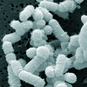
Research shows sharing of cavity-causing bacteria may not be only from mothers to children

New ongoing research from the University of Alabama at Birmingham Department of Biology and School of Dentistry is showing more evidence that children may receive oral microbes from other, nonrelative children. It was previously believed that these microbes were passed primarily from mother to child, but in a recent study presented at the American Society for Microbiology MICROBE 2016 Meeting in Boston, researchers found that 72 percent of children harbored at least one strain of the cavity-causing Streptococcus mutans not found in any cohabiting family members.

S. mutans is a bacterium that feeds on fermentable carbohydrates, in particular sucrose, that are frequently consumed by humans. After meals, S. mutans produces enamel-eroding acids, which makes S. mutans one of the main causes of tooth decay, or dental caries, in humans.

One hundred nineteen African-American children ages 12-18 months and 5-6 years who lived with at least one family member were a part of the study. The researchers collected samples from children periodically over the course of eight years. Momeni says that dental caries are more prevalent in minorities and low-socioeconomic groups.

"The literature tells us that we usually get this bacterium from our mothers," Momeni said. "This is because we most commonly have more interaction with our mothers when we are very young. However, our data supports that children who interact with other children at school or in nurseries can, and frequently do, contract this bacteria from each other." Momeni says any interaction that involves saliva, like sharing an ice cream cone or drinking after another child from the same cup or straw, can cause the microbes to be transferred.

Forty percent of the children in the study did not share any S. mutans strains with their mothers, and close to 20 percent of children shared these bacteria only with another child who lived in the household, such as a sibling or cousin. It is important to note that, for the strains of S. mutans not shared with anyone in the same household, approximately a third of the children had only a single isolate for a genotype, which could mean these rare strains may have nothing to do with the dental caries, and may be confounding the search for strains associated with the disease.

An interesting study that compared bacterial communities between healthy children and those that have a history of acute sinusitis (but not chronic sinusitis). The study specifically looked at the nasopharyngeal (NP)  microbiome (community of microbes) over the course of one year in the 2 groups of children, who were between the ages of 4 and 7. Nasopharyngeal pertains to the nose or nasal cavity and pharynx. They used modern methods of genetic analysis to test for bacterial species - and found a total of 951 species among the 47 children, of which 308 species had some "depletion" among those children with a history of sinusitis, and one species was increased in "abundance".

NP samples from children with a prior history of acute sinusitis were characterized by significant depletion of bacterial species, including those in the Akkermansia, Faecalibacterium prausnitzii, Clostridium, Lactobacillus, Prevotella, and Streptococcus species. But there was a siignificant increase "in relative abundance" in the bacterial species Moraxella nonliquefaciens. Once again, a study shows bacterial communities to be "out of whack" in those who've had sinusitis - this time in children. And the diminished diversity was linked to more frequent upper respiratory illnesses. The researchers mention the "possibility that the manipulation of the airway microbiota" could help prevent childhood respiratory diseases. Research by C.A. Santee et al from the Microbiome journal at BioMed Central:

Nasopharyngeal microbiota composition of children is related to the frequency of upper respiratory infection and acute sinusitis

Upper respiratory infections (URI) and their complications are a major healthcare burden for pediatric populations. Although the microbiology of the nasopharynx is an important determinant of the complications of URI, little is known of the nasopharyngeal (NP) microbiota of children, the factors that affect its composition, and its precise relationship with URI.

Healthy children (n = 47) aged 49–84 months from a prospective cohort study based in Wisconsin, USA, were examined. Demographic and clinical data and NP swab samples were obtained from participants upon entry to the study. All NP samples were profiled for bacterial microbiota using a phylogenetic microarray, and these data were related to demographic characteristics and upper respiratory health outcomes. The composition of the NP bacterial community of children was significantly related prior to the history of acute sinusitisHistory of acute sinusitis was associated with significant depletion in relative abundance of taxa including Faecalibacterium prausnitzii and Akkermansia spp. and enrichment of Moraxella nonliquefaciens. Enrichment of M. nonliquefaciens was also a characteristic of baseline NP samples of children who subsequently developed acute sinusitis over the 1-year study period. Time to develop URI was significantly positively correlated with NP diversity, and children who experienced more frequent URIs exhibited significantly diminished NP microbiota diversity (P ≤ 0.05). 

These preliminary data suggest that previous history of acute sinusitis influences the composition of the NP microbiota, characterized by a depletion in relative abundance of specific taxa. Diminished diversity was associated with more frequent URIs

....These observations indicate that the composition of the pediatric upper airway represents a critical factor that may either potentiate or protect against infection by respiratory pathogens. They also indicate that the interplay between the bacterial microbiota and respiratory pathogens associated with upper airway infection is important to consider.Both bacteria and viruses can influence each other’s pathogenicity [8] and a number of interactions between specific viruses and bacterial species have been reported in the airways [910]. For example, human rhinovirus infection was found to significantly increase the binding of Staphylococcus aureus, S. pneumoniae, or H. influenzae to primary human nasal epithelial cells [11]....

A total of 951 taxa were identified in baseline NP microbiota of participants (n = 47) in our cohort. These bacterial communities were variably composed of members of the Rickenellaceae, Lachnospiraceae, Verrucomicrobiaceae, Pseudomonadaceae, and Moraxellaceae as well as multiple unclassified members of the phylum Proteobacteria. .... Our study used independent NP samples collected from individual participants over a 12-month study period that spanned all four seasons. Season of sample collection also demonstrated a relationship with bacterial beta-diversity.

Compared with children who had no history of acute sinusitis (n = 33), those with a past history of acute sinusitis (n = 14) did not exhibit differences in α-diversity indices, suggesting that differences in microbiota characterizing these groups may be due to the enrichment or depletion of a subset of taxa within these bacterial communities. A total of 309 taxa (representing 101 genera) exhibited significant differences in relative abundance between children with and without a history of acute sinusitis. NP samples from children with a prior history of acute sinusitis were characterized by significant depletion of 308 of the 309 taxa, including those represented by Akkermansia, Faecalibacterium prausnitzii, Clostridium, Lactobacillus, Prevotella, and Streptococcus species. The only taxon that exhibited a significant increase in relative abundance in these subjects was represented by Moraxella nonliquefaciens. 

Children who experienced at least one URI (n = 17) within 60 days of collection of the baseline sample had significantly lower phylogenetic diversity compared to those who had no URIs within that time frame (n = 23). Time to development of URI, defined as the number of days between the collection of the baseline sample and the first incidence of URI (a value of 365 days was assigned to those children who did not experience a URI during the year of monitoring), was also significantly correlated with phylogenetic diversity .... Hence, these data indicate that diminished diversity of the NP microbiota is a precursor to URI in these children.  

In addition to Moraxella, a Corynebacterium was enriched in relative abundance in the NP microbiota of children who experienced acute sinusitis subsequent to baseline sample collection during the study period. ... However, Abreu et al. previously found Corynebacterium tuberculostearicum to be significantly enriched in the maxillary sinuses of adults with chronic rhinosinusitis compared to healthy control subjects [17]. The authors subsequently confirmed the ability of C. tuberculostearicum to induce acute sinusitis in the context of an antimicrobial-depleted murine model of sinus infection. Moreover co-installation of Lactobacillus sakei (one of a number of taxa acutely depleted in relative abundance among chronic rhinosinusitis patients) protected animals against C. tuberculostearicum infection [17]. Our pediatric data exhibits similarity with these murine studies, in that six members of the Lactobacillus genus were among those taxa most significantly depleted in relative abundance in the NP bacterial communities of children who developed sinusitis during our study. Five of these same taxa were also depleted in relative abundance in the NP microbial communities of children with a prior history of sinusitis. 

In addition to Lactobacillus, many other bacterial taxa including Akkermansia, Faecalibacterium prausnitzii, Clostridium, Prevotella, and Streptococcus species were depleted in relative abundance among children with a prior history of acute sinusitis. Though traditionally associated with gut microbiota, anaerobic bacterial species can exist in biofilms in the upper respiratory tract [18] and Akkermansia  and Faecalibacterium have previously been detected in the nasopharynx of children [1920]. While its role in the airway is unknown, gastrointestinal Akkermansia muciniphilia metabolizes mucin and has been shown to activate immune homeostasis, increasing host expression of antimicrobial peptides such as RegIIIγand improving barrier function via an increase in 2-oleoylgylcercerol [212223]. However, whether such mechanisms play a role at the airway mucosal surface remains to be determined. 

Mechanisms by which Lactobacillus and other bacterial species depleted in the NP microbiota of sinusitis patients may prevent the development of disease include competitive exclusion of pathogenic species. A previous murine study indicated that intra-nasal inoculation of mice with L. fermentum decreased S. pneumoniae burden throughout the respiratory tract and increased the number of activated macrophages in the lung and lymphocytes in the tracheal lamina propria [24]. Hence, it is plausible that the absence of NP genera with known competitive exclusion and immunomodulatory capabilities leads to pathogen expansion and associated clinical manifestations of upper airway infection. 

....We do show that a history of sinusitis, its pathophysiology or treatment, may shape the NP microbiota—which may inform future studies and their design. Additionally, though we recognize that the composition of the microbiota in the upper airways is likely highly influenced by antibiotic administration .... The pervasive effects of antimicrobials on the human microbiota are well-described [2627], and it is likely that lifetime antibiotic use plays an important role in shaping the baseline NP microbial community

The composition of the NP microbiota in healthy children between 49 and 84 months of age is associated with past and subsequent history of acute sinusitis and frequency of URI. Widespread bacterial taxon depletion and enrichment of M. liquefaciens and C. tuberculostearicum are associated with upper airway infection and the development of acute sinusitis. Collectively, these findings provide evidence of close connections between microbial colonization of the airways and susceptibility to upper respiratory illnesses in early childhood and raise the possibility that the manipulation of the airway microbiota could be applied to the prevention of childhood respiratory illnesses. 

Another view of  type 2 diabetes - that the gut microbiome is involved, specifically two gut bacteria: Prevotella copri and Bacteroides vulgatus. View them as the bad guys. The researchers point out "... the majority of overweight and obese individuals are insulin resistant and it is well known that dietary shifts to less calorie-dense eating and increased daily intake of any kind of vegetables and less intake of food rich in animal fat tend to normalize imbalances of gut microbiota and simultaneously improve insulin sensitivity of the host." In other words, eat more vegetables and fewer calories (if you're overweight or obese) to improve the gut microbes. This is similar to yesterday's post of research that viewed type 2 diabetes as "a response to overnutrition" and potentially reversible. From Medical Express:

Gut bacteria imbalance increases diabetes risk

Currently, scientists think the major contributors to insulin resistance are excess weight and physical inactivity, yet ground-breaking new research by an EU funded European-Chinese team of investigators called MetaHit have discovered that specific imbalances in the gut bacteria can cause insulin resistance, which confers an increased risk of health disorders like type 2 diabetes.

We show that specific imbalances in the gut microbiota are essential contributors to insulin resistance, a forerunner state of widespread disorders like type 2 diabetes, hypertension and atherosclerotic cardiovascular diseases, which are in epidemic growth," says Professor Oluf Pedersen, Metabolism Center, University of Copenhagen, and senior lead author of the paper.

In the Danish study of 277 non-diabetic individuals and 75 type 2 diabetic patients, there was close collaboration between the University of Copenhagen and the Technical University of Denmark with extensive international participation from a team of investigators, who performed analyses of the action of the insulin hormone. They monitored the concentrations of more than 1200 metabolites in blood and did advanced DNA-based studies of hundreds of bacteria in the human intestinal tract to explore if certain imbalances in gut microbiota are involved in the causation of common metabolic and cardiovascular disorders.

The researchers observed that people who had a decreased capacity of insulin action, and therefore were insulin resistant, had elevated blood levels of a subgroup of amino acids called branched-chain amino acids (BCAAs). Importantly, the rise of BCAAs levels in blood was related to specific changes in the gut microbiota composition and function.

The main drivers behind the gut bacterial biosynthesis of BCAAs turned out to be the two bacteria Prevotella copri and Bacteroides vulgatus. To test mechanistically if gut bacteria were a true cause of insulin resistance, the researchers fed mice with the Prevotella copri bacteria for 3 weeks. Compared with sham fed mice the Prevotella copi fed mice developed increased blood levels of BCAAs, insulin resistance and intolerance to glucose.

"Most people with insulin resistance do not know that they have it. However, it is known that the majority of overweight and obese individuals are insulin resistant and it is well known that dietary shifts to less calorie-dense eating and increased daily intake of any kind of vegetables and less intake of food rich in animal fat tend to normalize imbalances of gut microbiota and simultaneously improve insulin sensitivity of the host," adds Pedersen. (Original study)

Newly published research found that children who are thumb-suckers or nail-biters are less likely to develop atopic sensitization or allergic sensitivities (as measured by positive skin-prick tests to common allergens). And, if they have both 'habits', they are even less likely to be allergic to such things as house dust mites, grass, cats, dogs, horses, wool, or airborne fungi. The finding emerges from a longitudinal study which followed the progress of 1,037 persons born in Dunedin, New Zealand in 1972-1973 from childhood into adulthood. However, the researchers found no relationship to these 2 habits to allergic asthma or "hay fever" - a contradictory finding that the researchers don't have an answer for.

"Our findings are consistent with the hygiene theory that early exposure to dirt or germs reduces the risk of developing allergies," said Professor Sears (one of the researchers).  The researchers were testing the idea that the common childhood habits of thumb-sucking and nail-biting would increase microbial exposures, affecting the immune system and reducing the development of allergic reactions also known as atopic sensitization. 31% of the children were frequent thumb suckers or nail biters.

Among all children at 13 years old, 45% showed atopic sensitization, but among those with no habits 49% had allergic sensitization; and those with one oral habit - 40% had allergic sensitization. Among those with both habits, only 31% had allergic sensitization. This trend continued into adulthood, and showed no difference depending on smoking in the household, ownership of cats or dogs; or exposure to house dust mites.

Excerpts of the study from Pediatrics: Thumb-Sucking, Nail-Biting, and Atopic Sensitization, Asthma, and Hay Fever

The hygiene hypothesis suggests that early-life exposure to microbial organisms reduces the risk of developing allergies. Thumb-sucking and nail-biting are common childhood habits that may increase microbial exposures. We tested the hypothesis that children who suck their thumbs or bite their nails have a lower risk of developing atopy, asthma, and hay fever in a population-based birth cohort followed to adulthood. Parents reported children’s thumb-sucking and nail-biting habits when their children were ages 5, 7, 9, and 11 years. Atopic sensitization was defined as a positive skin-prick test (≥2-mm weal) to ≥1 common allergen at 13 and 32 years. 

Thirty-one percent of children were frequent thumb-suckers or nail-biters at ≥1 of the ages. These children had a lower risk of atopic sensitization at age 13 years  and age 32 years. These associations persisted when adjusted for multiple confounding factors. Children who had both habits had a lower risk of atopic sensitization than those who had only 1. No associations were found for nail-biting, thumb-sucking, and asthma or hay fever at either age.

What This Study Adds: Children who sucked their thumbs or bit their nails between ages 5 and 11 years were less likely to have atopic sensitization at age 13. This reduced risk persisted until adulthood. There was no association with asthma or hay fever.

The “hygiene hypothesis” was suggested by Strachan1 to explain why children from larger families and those with older siblings are less likely to develop hay fever. Strahan hypothesized that this could be explained if “allergic diseases were prevented by infection in early childhood transmitted by unhygienic contact with older siblings, or acquired prenatally from a mother infected by contact with her older children.” The hypothesis is supported by evidence showing that children who grow up in large families are at greater risk of coming into contact with more infections....The hygiene hypothesis remains controversial, however, as it is unable to fully explain many associations, including the rise of allergies in “unhygienic” inner-city environments, and why probiotics are ineffective at preventing allergic diseases.3

Thumb-sucking and nail-biting are common oral habits among children, although the reported prevalence varies widely, from <1% to 25%.47 These habits have the potential to increase the exposure to environmental microorganisms, and have been associated with the oral carriage of Enterobacteriaceae, such as Escherichia coli and intestinal parasite infections.812 It seems likely that thumb-sucking and nail-biting would introduce a wide variety of microbes into the body, thus increasing the diversity of the child’s microbiome. If the hygiene hypothesis is correct, it is plausible that this would influence the risk for allergies.... 

Of 1013 children providing data, 317 (31%) had ≥1 oral habit: there was no significant sex difference in prevalence of these habits. Of the 724 children who had skin-prick tests at age 13 years, 328 (45%) showed atopic sensitization. The prevalence of sensitization was lower among children who had an oral habit (38%) compared with those who did not (49%) (P = .009). The lower risk of atopic sensitization was similar for thumb-sucking and nail-biting. Children with only 1 habit were less likely to be atopic (40%) than children with no habit at all (49%), but those with both habits had the lowest prevalence of sensitization (31%) .