 Interesting new research found health benefits to the brain from daily low intake of alcohol (equivalent to about 2 1/2 drinks per day). The University of Rochester (in New York) researchers found that while low daily (chronic) levels of alcohol were beneficial to the brain's glymphatic system, higher daily levels or binge drinking was not. And the low daily levels of alcohol intake was also better for the glymphatic system than no alcohol at all (the control group). In 2015 this same research team described the glymphatic system as not just the brain’s “waste-clearance system,” but as potentially helping fuel the brain by transporting glucose, lipids, amino acids, and neurotransmitters.
I'm sure this study will be greeted by many as great news, but remember it was done with MICE, and not humans, so one should be cautious in generalizing the results. But the researchers think that it does apply to humans, and may explain why some studies find some health benefits to low levels of daily alcohol intake, even better than no alcohol, and many negative effects to higher levels of alcohol intake - thus the J-shaped curve of effects seen in studies. [NOTE: Studies also find that alcohol consumption can cause cancer, and this is dose related. Studies find the Mediterranean diet (which includes low to moderate levels of alcohol) beneficial for brain health.]
By the way - no, the mice didn't receive wine as the press release from the Univ. of Rochester says. The mice actually received "intraperitoneal injections of low, intermediate, and high doses of ethanol" or just plain saline (the control group). From Science Daily:
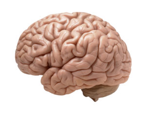
In wine, there's health: Low levels of alcohol good for the brain
While a couple of glasses of wine can help clear the mind after a busy day, new research shows that it may actually help clean the mind as well. The new study, which appears in the journal Scientific Reports, shows that low levels of alcohol consumption tamp down inflammation and helps the brain clear away toxins, including those associated with Alzheimer's disease.
"Prolonged intake of excessive amounts of ethanol is known to have adverse effects on the central nervous system," said Maiken Nedergaard, M.D., D.M.Sc., co-director of the Center for Translational Neuromedicine at the University of Rochester Medical Center (URMC) and lead author of the study. "However, in this study we have shown for the first time that low doses of alcohol are potentially beneficial to brain health, namely it improves the brain's ability to remove waste." The finding adds to a growing body of research that point to the health benefits of low doses of alcohol. While excessive consumption of alcohol is a well-documented health hazard, many studies have linked lower levels of drinking with a reduced risk of cardiovascular diseases as well as a number of cancers.
Nedergaard's research focuses on the glymphatic system, the brain's unique cleaning process that was first described by Nedergaard and her colleagues in 2012. They showed how cerebral spinal fluid (CSF) is pumped into brain tissue and flushes away waste, including the proteins beta amyloid and tau that are associated with Alzheimer's disease and other forms of dementia. Subsequent research has shown that the glymphatic system is more active while we sleep, can be damaged by stroke and trauma, and improves with exercise.
The new study, which was conducted in mice, looked at the impact of both acute and chronic alcohol exposure. When they studied the brains of animals exposed to high levels of alcohol over a long period of time, the researchers observed high levels of a molecular marker for inflammation, particularly in cells called astrocytes which are key regulators of the glymphatic system. They also noted impairment of the animal's cognitive abilities and motor skills.
Animals that were exposed to low levels of alcohol consumption, analogous to approximately 2 ½ drinks per day, actually showed less inflammation in the brain and their glymphatic system was more efficient in moving CSF through the brain and removing waste, compared to control mice who were not exposed to alcohol. The low dose animals' performance in the cognitive and motor tests was identical to the controls.
"The data on the effects of alcohol on the glymphatic system seemingly matches the J-shaped model relating to the dose effects of alcohol on general health and mortality, whereby low doses of alcohol are beneficial, while excessive consumption is detrimental to overall health" said Nedergaard. "Studies have shown that low-to-moderate alcohol intake is associated with a lesser risk of dementia, while heavy drinking for many years confers an increased risk of cognitive decline. This study may help explain why this occurs. Specifically, low doses of alcohol appear to improve overall brain health." [Original study. Especially interesting is the Introduction & Discussion sections.]
 This is a topic that is totally neglected: What will it feel like when the carbon dioxide levels in the atmosphere (the air) increase as our climate changes? What kinds of effects will it have on our thought processes (our cognition)? The reason I mention this is because research shows that as carbon dioxide (CO2) levels increase in rooms with people in them, it feels "stuffy" and people's thinking (cognitive processes) deteriorate. They don't think and work as effectively. Air starts to feel "stuffy" at about 600 ppm (parts per million). Our current CO2 levels in outside air are already above 400 ppm, and the levels are forecast to keep rising.
 A number of recent studies and articles have discussed the effectiveness of diet in treating or preventing depression with the main conclusion that yes, it helps. Now an observational study (that will be presented in April) found that elderly people following the DASH diet most closely were 11% less likely to become depressed over time than those that did not.
 The spice turmeric is a very popular supplement nowadays, believed to have all sorts of health benefits due to the curcumin in it (e.g. that it is anticancer, anti-Alzheimer's, anti inflammatory). And yes, studies in the lab (in vitro and in vivo) look very promising. However, a large 2017