Skip to content

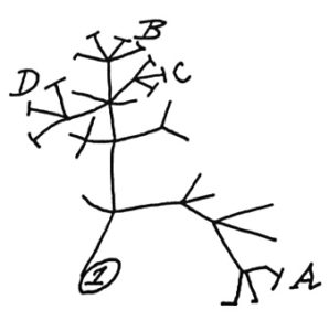
In 1837, Charles Darwin sketched a simple tree of life (shown left) to illustrate the idea that all living things share a common ancestor. Ever since then, scientists have been adding names to the tree of life, including a massive effort of 2.3 million named species of animals, plants, fungi and microbes in 2015. A tree of life is a visual hypothesis of how scientists think species are related to one another, so it has been evolving over the years as more information is learned and species discovered.

Now a group of 17 researchers have sketched out a radically different tree of life. It has two main trunks—one full of bacteria and another comprised of archaea, a group of single-celled microbes that run on very different biochemistry. The eukaryotes—the domain that includes all animals, fungi, and plants—are crowded on a thin branch coming off the archaeal trunk. About half of these bacterial branches belong to a supergroup, which was discovered very recently and is currently known as the candidate phyla radiation. Within these branches are numerous species that we’re almost completely ignorant about, and they’ve never been isolated or grown in a lab (with one exception called TM7). In fact, this supergroup of bacteria and “other lineages that lack isolated representatives clearly comprise the majority of life’s current diversity,” write researchers Hug and Banfield. Wow. From Science Daily:

Wealth of unsuspected new microbes expands tree of life

Scientists have dramatically expanded the tree of life, which depicts the variety and evolution of life on Earth, to account for over a thousand new microscopic life forms discovered over the past 15 years. The expanded view finally gives bacteria and Archaea their due, showing that about two-thirds of all diversity on Earth is bacterial -- half bacteria that cannot be isolated and grown in the lab -- while nearly one-third is Archaeal.

Much of this microbial diversity remained hidden until the genome revolution allowed researchers like Banfield to search directly for their genomes in the environment, rather than trying to culture them in a lab dish. Many of the microbes cannot be isolated and cultured because they cannot live on their own: they must beg, borrow or steal stuff from other animals or microbes, either as parasites, symbiotic organisms or scavengers.

The new tree, to be published online April 11 in the new journal Nature Microbiology, reinforces once again that the life we see around us -- plants, animals, humans and other so-called eukaryotes -- represent a tiny percentage of the world's biodiversity.

"Bacteria and Archaea from major lineages completely lacking isolated representatives comprise the majority of life's diversity," said Banfield.....According to first author Laura Hug,... the more than 1,000 newly reported organisms appearing on the revised tree are from a range of environments, including a hot spring in Yellowstone National Park, a salt flat in Chile's Atacama desert, terrestrial and wetland sediments, a sparkling water geyser, meadow soil and the inside of a dolphin's mouth. All of these newly recognized organisms are known only from their genomes.

One striking aspect of the new tree of life is that a group of bacteria described as the "candidate phyla radiation" forms a very major branch. Only recognized recently, and seemingly comprised only of bacteria with symbiotic lifestyles, the candidate phyla radiation now appears to contain around half of all bacterial evolutionary diversity.

Charles Darwin first sketched a tree of life in 1837 as he sought ways of showing how plants, animals and bacteria are related to one another. The idea took root in the 19th century, with the tips of the twigs representing life on Earth today, while the branches connecting them to the trunk implied evolutionary relationships among these creatures.....Archaea were first added in 1977 after work showing that they are distinctly different from bacteria, though they are single-celled like bacteria. A tree published in 1990 by microbiologist Carl Woese was "a transformative visualization of the tree," Banfield said. With its three domains, it remains the most recognizable today.

With the increasing ease of DNA sequencing in the 2000s, Banfield and others began sequencing whole communities of organisms at once and picking out the individual groups based on their genes alone. This metagenomic sequencing revealed whole new groups of bacteria and Archaea, many of them from extreme environments, such as the toxic puddles in abandoned mines, the dirt under toxic waste sites and the human gut. Some of these had been detected before, but nothing was known about them because they wouldn't survive when isolated in a lab dish.

For the new paper, Banfield and Hug teamed up with more than a dozen other researchers who have sequenced new microbial species, gathering 1,011 previously unpublished genomes to add to already known genome sequences of organisms representing the major families of life on Earth.....The analysis, representing the total diversity among all sequenced genomes, produced a tree with branches dominated by bacteria, especially by uncultivated bacteria. A second view of the tree grouped organisms by their evolutionary distance from one another rather than current taxonomic definitions, making clear that about one-third of all biodiversity comes from bacteria, one-third from uncultivable bacteria and a bit less than one-third from Archaea and eukaryotes. (Original article and diagrams.)

This is a new and expanded view of the tree of life, with clusters of bacteria (left), uncultivable bacteria called 'candidate phyla radiation' (center, purple) and, at lower right, the Archaea and eukaryotes (green), including humans.
Credit: Graphic by Zosia Rostomian, Lawrence Berkeley National Laboratory

Another article commenting that increasing the amount of dietary fiber eaten by people eating a typical Western diet (which is low in fiber) will improve their gut microbiome (community of microbes). Research is finding that the added dietary fiber is food (nutrition) for microbes in the gut, and eating additional fiber daily will help restore or increase bacterial diversity, which then should lead to health benefits. Note: Easy ways to increase dietary fiber are increasing intake of whole grains, legumes, nuts, seeds, fruits, and vegetables. Think of food writer Michael Pollan's advice: "Eat food. Not too much. Mostly plants."

Researchers feel that fiber intake needs to be increased to more than current dietary guidelines, and that beneficial effects to the microbiome starts to occur rapidly (within 2 weeks) of changing to a higher fiber diet. This post from January 21, 2016 discussed the Sonnenburg research on gut microbe depletion (from a low fiber diet), and this April 28, 2015 post discussed the O'Keefe research (changing the diet has big effect on colon cancer risk) - both studies are mentioned below. See the page Feeding Your Gut Microbes for more information. From Science Daily:

Can more fiber restore microbiome diversity?

Scientists are pushing to restore human health in Western countries by changing our diet to restore the microbial species lost over the evolution of Western diet. Researchers advocate for strategically increasing dietary fiber intake as one path forward in regaining microbial biodiversity.

Insufficient nutrients for our gut microbes have been linked to a loss of certain beneficial bacterial species in industrialized societies and are likely impacting our immunological and metabolic health, although more data is needed. For example, most Westerners consume half of the amount of dietary fiber recommended by dietary guidelines, which nutritionists refer to as the "fiber gap," which is a problem because dietary fiber is the primary source of nutrition (e.g., carbohydrates) accessible to gut bacteria in humans.

"The idea to boost fiber levels is not new," says Jens Walter of the University of Alberta, Canada. "However, depletion of the microbiome adds a new perspective to this low-fiber Western diet that we are currently eating." Earlier this year, Stanford University's Justin Sonnenburg found that mice fed a typical Western diet (high in fat and carbohydrates and low in fiber) transferred a lower diversity of beneficial microbial species to future generations. The re-introduction of the microbes' preferred fiber at that stage did not result in a return of some (good) species, indicating that extinctions had occurred in only a few generations.

Walter and co-author Edward Deehan, his PhD student, are concerned that a dramatic shift away from a diet similar to the one under which the human-microbiome symbiosis evolved is a key factor in the rise of non-communicable disorders like obesity. "There is a lot of epidemiological evidence that fiber is beneficial, and food products containing dietary fiber have FDA-approved health claims for both colon cancer and coronary heart disease. There is also quite a bit of clinical evidence (although it is less consistent)," Walter says. "The most pressing issue at the moment that neither consumption of fiber in society nor the doses used in clinical research are high enough."

People living in non-industrialized societies have an average intake of fiber that is much higher than the low norms of Western societies. The authors note the recent work from the Stephen J.D. O'Keefe lab in Nature Communications in which modern African-Americans were given a traditional South-African diet that contained 55 grams of daily dietary fiber and had improved markers for colon cancer within two weeks.

 Numerous studies link toxoplasmosis to a number of psychiatric conditions, including bipolar disorder, schizophrenia, and obsessive-compulsive disorder. This infection is also linked to aspects of behavior and personality, from impulsivity to prolonged feelings of guilt and excessive worry, and may even slow a person's reflexes. Now new research has linked latent toxoplasmosis infection to aggression and impulsivity. Those with a psychiatric condition called intermittent explosive disorder (IED) have a greater likelihood of toxoplasmosis infection (when compared to people without IED). People who have IED typically experience recurrent outbursts of extreme, impulsive anger, such as seen in road rage.

Three groups (normal healthy control group, intermittent explosive disorder (IED) group, and non-IED psychiatric disorders group) were studied, and in all 3 groups those who tested positive for the infection had higher levels of aggression within their group. 22% of the IED group had the infection, 17% in the non-IED psychiatric group, and only 9% in the healthy normal control group.This research suggested that toxoplasmosis infection is linked to aggression and impulsivity (with aggression being stronger).

Toxoplasmosis is caused by an infection with Toxoplasma gondii, a protozoan parasite carried by cats. It can also infect humans, through contact with cat feces, poorly cooked meat or contaminated water, and as many as one-third of the world’s population may be infected. The person may not feel sick, but the parasite may form cysts in the brain where it can remain for the rest of a person’s life. In this study they determined that someone had a latent T. gondii infection by the circulating immunoglobulin G (IgG) antibodies to T. gondii in the blood (IgG > 12 IU). And yes, the infection can be successfully treated with medications. From Science Daily:

People with rage disorder twice as likely to have latent toxoplasmosis parasite infection

Individuals with a psychiatric disorder involving recurrent bouts of extreme, impulsive anger--road rage, for example--are more than twice as likely to have been exposed to a common parasite than healthy individuals with no psychiatric diagnosis. In a study involving 358 adult subjects, a team led by researchers from the University of Chicago found that toxoplasmosis, a relatively harmless parasitic infection carried by an estimated 30 percent of all humans, is associated with intermittent explosive disorder and increased aggression.

"Our work suggests that latent infection with the toxoplasma gondii parasite may change brain chemistry in a fashion that increases the risk of aggressive behavior," said senior study author Emil Coccaro, MD, Ellen. C. Manning Professor and Chair of Psychiatry and Behavioral Neuroscience at the University of Chicago. "However, we do not know if this relationship is causal, and not everyone that tests positive for toxoplasmosis will have aggression issues," Coccaro said, adding that additional studies are needed.

Intermittent explosive disorder (IED) is defined by the Diagnostic and Statistical Manual of Mental Disorders, Fifth Edition, as recurrent, impulsive, problematic outbursts of verbal or physical aggression that are disproportionate to the situations that trigger them. IED is thought to affect as many as 16 million Americans, more than bipolar disorder and schizophrenia combined.....examined possible connections to toxoplasmosis, an extremely common parasitic infection. Transmitted through the feces of infected cats, undercooked meat or contaminated water, toxoplasmosis is typically latent and harmless for healthy adults. However, it is known to reside in brain tissue, and has been linked to several psychiatric diseases, including schizophrenia, bipolar disorder and suicidal behavior.

The research team recruited 358 adult subjects from the U.S., who were evaluated for IED, personality disorder, depression and other psychiatric disorders. Study participants were also scored on traits including anger, aggression and impulsivity. Participants fell into one of three groups. Roughly one third had IED. One third were healthy controls with no psychiatric history. The remaining third were individuals diagnosed with some psychiatric disorder, but not IED. This last group served as a control to distinguish IED from possible confounding psychiatric factors.

The research team found that IED-diagnosed group was more than twice as likely to test positive for toxoplasmosis exposure (22 percent) as measured by a blood test, compared to the healthy control group (9 percent). Around 16 percent of the psychiatric control group tested positive for toxoplasmosis, but had similar aggression and impulsivity scores to the healthy control group. IED-diagnosed subjects scored much higher on both measures than either control group.

Across all study subjects, toxoplasmosis-positive individuals scored significantly higher on scores of anger and aggression. The team noted a link between toxoplasmosis and increased impulsivity, but when adjusted for aggression scores, this link became non-significant. This finding suggests toxoplasmosis and aggression are most strongly correlated. However, the authors caution that the study results do not address whether toxoplasmosis infection may cause increased aggression or IED. "Correlation is not causation, and this is definitely not a sign that people should get rid of their cats," said study co-author Royce Lee, MD.... "We don't yet understand the mechanisms involved..."

 Toxoplasma gondii  tissue cyst in a mouse brain Credit: Jitinder P. Dubey, Wikipedia

The eye has a normal community of microbes or eye microbiome, just like other body sites (i.e., the gut, the sinuses, the mouth). This community of bacteria is thought to offer resistance from invaders (such as pathogenic bacteria).

The researchers of a recent study found differences in the eye microbiome of contact lens wearers as compared to non-lens wearers. The results indicate that wearing contact lenses "alters the microbial structure of the ocular conjunctiva, making it more similar to that of the skin microbiota" (the community of microbes living on the skin).

Further research is needed to determine whether these differences in the eye microbiome in contact lens wearers is the reason why contact lens wearers develop more eye conditions and infections (such as giant papillary conjunctivitis and keratitis). Over 30 million Americans wear contact lenses so these are important issues.

From Science Daily: Contact lenses alter eye bacteria, making it more skin-like

Contact lenses may alter the natural microbial community of the eyes, according to a study published this week in mBio®, an online open-access journal of the American Society for Microbiology. In a study of 58 adults seeking outpatient eye care, researchers at New York University School of Medicine found that contact lenses make the eye microbiome more skin-like, with higher proportions of the skin bacteria Pseudomonas, Acinetobacter, Methylobacterium, and Lactobacillus and lower proportions of Haemophilus, Streptococcus, Staphylococcus, and Corynebacterium.

It's unclear how these changes occur, said senior study author Maria Dominguez-Bello, PhD, an associate professor of medicine at the university, "if these bacteria are transferred from the fingers to the lens and to the eye surface, or if the lenses exert selective pressures on the eye bacterial community in favor of skin bacteria." Wearing contact lenses has been identified as a risk factor for the development of eye infections such as giant papillary conjunctivitis and keratitis, "so these questions are important," she said.

Researchers used a laboratory technique called 16s rRNA sequencing to compare the bacterial communities of the conjunctiva (the eye surface) and the skin under the eye from 58 adults. They also analyzed samples from 20 of the study participants (9 lens wearers and 11 non-lens wearers) at three time points over the course of six weeks....researchers found a higher diversity of bacteria on the ocular surface than on the skin under the eye or on the contact lenses, which was a surprising result, Dominguez-Bello said.

The ocular surface microbiota of those who wore contact lenses was more skin-like compared to those who did not wear lenses. It was enriched in the bacteria Pseudomonas, Acinetobacter, Methylobacterium, and Lactobacillus. In non-lens wearers, these bacteria were detected at a higher relative abundance in skin samples compared to the eye (except for Lactobacillus), suggesting that these bacteria could be classified as skin bacteria. The bacteria Haemophilus, Streptococcus, Staphylococcus, and Corynebacterium were depleted in the ocular microbiota of lens wearers compared to non-lens wearers.

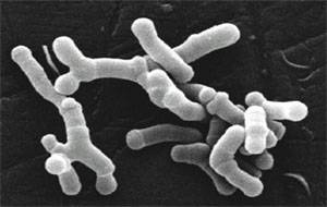
The possibility of giving microbes in the future (whether bacteria, viruses, or fungi) to treat cancer is amazing. Of course big pharma is pursuing this line of research, which is called immunotherapy (stimulating the body's ability to fight tumors). The Bloomberg Business article discusses a number of big pharma companies entering the field and their main focus. The study in the journal Science finding that giving common beneficial bacteria (Bifidobacterium breve and Bifidobacterium longum) to mice to slow down melanoma tumor growth is a first step. The researchers themselves said that the 2 common beneficial bacteria species exhibited anti-tumor activity in the mice and was as effective as an immunotherapy in controlling the growth of skin cancer. But note that the bacteria needed to be live. Stay tuned....

From Bloomberg News: How Gut Bacteria Are Shaking Up Cancer Research

Top scientists at Roche Holding AG and AstraZeneca Plc are sizing up potential allies in the fight against cancer: the trillions of bacteria that live in the human body. "Five years ago, if you had asked me about bacteria in your gut playing an important role in your systemic immune response, I probably would have laughed it off," Daniel Chen, head of cancer immunotherapy research at Roche’s Genentech division, said in a phone interview. "Most of us immunologists now believe that there really is an important interaction there."

Two recent studies published in the journal Science have intrigued Chen and others who are developing medicines called immunotherapies that stimulate the body’s ability to fight tumors.In November, University of Chicago researchers wrote that giving mice Bifidobacterium, which normally resides in the gastrointestinal tract, was as effective as an immunotherapy in controlling the growth of skin cancer. Combining the two practically eliminated tumor growth. In the second study, scientists in France found that some bacterial species activated a response to immunotherapy, which didn’t occur without the microbes.  ...continue reading "Research Using Bacteria to Treat Cancer"

A thought-provoking article by Heiman and Greenway was just published in the journal Molecular Metabolism making the case that changes in farming practices over the last 50 years have resulted in decreased agricultural diversity which, in turn, has resulted in decreased dietary diversity, and that the reduction in dietary diversity has changed and decreased the richness of the human gut microbiota (microbes living in the gut). And meanwhile, during the past 50 years, the rates of obesity, type 2 diabetes, and inflammatory bowel diseases sharply increased - and in each of these conditions there is a reduction of the gut microbial diversity. Similar views have also been stated by others in the field of microbiology.

The thinking is that the more diverse the diet, the more diverse the gut microbiome (and healthier), and the more it can adapt to disturbances. Heiman and Greenway state: "Unfortunately, dietary diversity has been lost during the past 50 years because of economic pressures for greater food production to support a growing world population.... Of the 250,000 to 300,000 known edible plant species, humans use only 150 to 200...Today, 75 percent of the world's food is generated from only 12 plants and five animal species."

Also, agricultural practices of using antibiotics as growth promoters for poultry, swine, and cattle further harm the human gut microbiome when the meat is ingested by humans, and pesticide residues on crops ingested by humans may have gut microbiome effects. Even emulsifiers, used in processed foods, reduce microbial richness. Every time a person goes on a certain diet (vegan, Paleo, etc) or makes dietary choices in which some foods are eliminated, it makes it easier for some microbial species, and gives them a competitive advantage over other gut microbes. From Science Daily:

Reduction in dietary diversity impacts richness of human gut microbiota

Changes in farming practices over the last 50 years have resulted in decreased agro-diversity which, in turn, has resulted in decreased dietary diversity. The significant impact of this change in dietary richness on human health is an emerging topic for discussion

Heiman and Greenway describe how the reduction in dietary diversity has changed the richness of human gut microbiota, the community of microorganisms living in the gut. The researchers point out that healthy individuals have diverse gut microbiota and many of the common pathologies of the 21st century, including type 2 diabetes, obesity and inflammatory bowel disease, are associated with reduced microbiotic richness.

Gut microbiota function as an endocrine organ, metabolizing specific nutrients from the diet and producing specific substances that act as metabolic signals in the host. It follows then that highly specialized diets will change the landscape of the gut microbiome over time. In fact, it takes only a few days of changing diet to alter the microbiotic makeup of the human gut. And if the dietary change involves elimination of one or more macronutrients (think Atkins or Paleo or vegan), humans are essentially selecting for some microbiotic species over others.

The importance of microbiota diversity cannot be overstated. They produce an abundance of important molecules for the host and with increased variation comes increased adaptability and an increased range of physiological responses. "The greater the repertoire of signals, the more likely is the ability to maintain homeostasis when dietary intake is perturbed," explain Heiman and Greenway. "Furthermore, because each particular macronutrient has the potential to be metabolized by microbiota into unique metabolic signals, the greater the variety in signals, the greater the variety of responses possible."

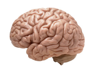
 Two articles about the link between Alzheimer's disease (AD) and microbes this past week: a study linking periodontal disease and Alzheimer's, and the other a journal editorial (written by an international team of 31 researchers) suggesting that we need to more closely look at the role of microbes in Alzheimer's disease, especially herpes virus, chlamydia and spirochaete bacteria.

This team is suggesting an "infectious cause" for Alzheimer's, an example being the reactivation of herpes simplex virus type 1 (HSV1) in the person. The researchers state that "regarding HSV1, about 100 publications by many groups indicate directly or indirectly that this virus is a major factor in the disease".  The team also mentions the possibility of fungi infection in some cases (see my November 6, 2015 post about a study finding fungal involvement). Both articles mention that treatment of the diseases with some form of antimicrobials or antivirals could possibly treat Alzheimer's disease, and that trails now need to be done.

From Science Daily: Link between gum disease and cognitive decline in Alzheimer’s

A new study has found a link between gum disease and greater rates of cognitive decline in people with early stages of Alzheimer's Disease. Periodontitis or gum disease is common in older people and may become more common in Alzheimer's disease because of a reduced ability to take care of oral hygiene as the disease progresses. Higher levels of antibodies to periodontal bacteria are associated with an increase in levels of inflammatory molecules elsewhere in the body, which in turn has been linked to greater rates of cognitive decline in Alzheimer's disease in previous studies.

The presence of gum disease at baseline was associated with a six-fold increase in the rate of cognitive decline in participants over the six-month follow-up period of the study. Periodontitis at baseline was also associated with a relative increase in the pro-inflammatory state over the six-month follow-up period. The authors conclude that gum disease is associated with an increase in cognitive decline in Alzheimer's Disease, possibly via mechanisms linked to the body's inflammatory response.....However, growing evidence from a number of studies links the body's inflammatory response to increased rates of cognitive decline, suggesting that it would be worth exploring whether the treatment of gum disease might also benefit the treatment of dementia and Alzheimer's Disease.

From Journal of Alzheimer's Disease: Microbes and Alzheimer’s Disease

We are researchers and clinicians working on Alzheimer’s disease (AD) or related topics, and we write to express our concern that one particular aspect of the disease has been neglected, even though treatment based on it might slow or arrest AD progression. We refer to the many studies, mainly on humans, implicating specific microbes in the elderly brain, notably herpes simplex virus type 1 (HSV1), Chlamydia pneumoniae, and several types of spirochaete, in the etiology of AD [1–4]. Fungal infection of AD brain [5, 6] has also been described, as well as abnormal microbiota in AD patient blood [7]. The first observations of HSV1 in AD brain were reported almost three decades ago [8]. The ever-increasing number of these studies (now about 100 on HSV1 alone) warrants re-evaluation of the infection and AD concept.

AD is associated with neuronal loss and progressive synaptic dysfunction, accompanied by the deposition of amyloid-β (Aβ) peptide, a cleavage product of the amyloid-β protein precursor (AβPP), and abnormal forms of tau protein, markers that have been used as diagnostic criteria for the disease [9, 10]. These constitute the hallmarks of AD, but whether they are causes of AD or consequences is unknown. We suggest that these are indicators of an infectious etiology. In the case of AD, it is often not realized that microbes can cause chronic as well as acute diseases; that some microbes can remain latent in the body with the potential for reactivation, the effects of which might occur years after initial infection; and that people can be infected but not necessarily affected, such that ‘controls’, even if infected, are asymptomatic [2].

Regarding HSV1, about 100 publications by many groups indicate directly or indirectly that this virus is a major factor in the disease....The only opposing reports, two not detecting HSV1 DNA in elderly brains and another not finding an HSV1–APOE association, were published over a decade ago [76–78]. However, despite all the supportive evidence, the topic is often dismissed as ‘controversial’. One recalls the widespread opposition initially to data showing that viruses cause some types of cancer, and that a bacterium causes stomach ulcers.

In summary, we propose that infectious agents, including HSV1, Chlamydia pneumonia, and spirochetes, reach the CNS and remain there in latent form. These agents can undergo reactivation in the brain during aging, as the immune system declines, and during different types of stress (which similarly reactivate HSV1 in the periphery). The consequent neuronal damage— caused by direct viral action and by virus-induced inflammation— occurs recurrently, leading to (or acting as a cofactor for) progressive synaptic dysfunction, neuronal loss, and ultimately AD.

AD causes great emotional and physical harm to sufferers and their carers, as well as having enormously damaging economic consequences. Given the failure of the 413 trials of other types of therapy for AD carried out in the period 2002–2012 [79], antiviral/antimicrobial treatment of AD patients, notably those who are APOE ɛ 4 carriers, could rectify the ‘no drug works’ impasse. We propose that further research on the role of infectious agents in AD causation, including prospective trials of antimicrobial therapy, is now justified.

 Lyme disease is caused by the bacterium Borrelia burgdorferi and is typically treated with antibiotics. This study may go a long way in explaining why some people do not seem to respond to Lyme disease treatment, and why they continue to feel sick even after prolonged antibiotic therapy. The researchers discussed how, in addition to the familiar spirochete form, B. burgdorferi can transform from spirochetes into round body forms in the presence of various unfavorable environmental conditions, including the presence of antimicrobial agents (antibiotics). And that the different forms respond to different antibiotic treatments!

But now they found that this bacterium has an additional form, which they refer to as biofilm, and which may be resistant to even very aggressive antibiotic (antimicrobial) treatments. They say this is the first study that demonstrates the presence of Borrelia biofilm in infected human skin tissues. From Medical Xpress:

Lyme disease 'Biofilm' eludes antibiotics: report

In many cases, Lyme disease returns after a patient has completed antibiotic treatment, and this finding may help explain why that occurs, the researchers said. University of New Haven researchers determined that Lyme disease-causing Borrelia burgdorferi bacteria produces a biofilm that makes it up to 1,000 times more resistant to antibiotics than other bacteria.

The discovery may lead to new ways to treat Lyme disease, said study author Eva Sapi, head of biology and environmental sciences at the university. "These findings could change the way we think about Lyme disease, especially in patients where it seems to be a persistent disease, despite long-term antibiotic treatment," she said in a news release from the Connecticut-based university.

"This recent finding could help to better understand how Borrelia can survive treatment and ... will provide novel therapeutic targets for chronic Lyme disease, with the hope of eradicating Borrelia in these patients," Sapi added. (original study)

 Borrelia burgdorferi  Credit: CDC

Very exciting research IF it pans out - the idea of treating (some) cancers with probiotics (beneficial bacteria). This study was done on mice, and some mice started the probiotic mixture one week before they gave the mice the liver cancer, so...more limitations there. But the idea is so tantalizing and wonderful... And what was in the mixture of bacteria (called probiotic Prohep) that the mice ate that had beneficial results of shrinking liver tumors? The probiotic Prohep is composed of Lactobacillus rhamnosus GG (LGG), Escherichia coli Nissle 1917 (EcN), and heat inactivated VSL#3 (1:1:1).  VSL#3 contains: Streptococcus thermophilus, Bifidobacterium breve, Bifidobacterium longum, Bifidobacterium infantis, Lactobacillus acidophilus, Lactobacillus plantarum, Lactobacillus paracasei, and Lactobacillus delbrueckii. Note that Lactobacillus rhamnosus and some of the others are already found in many probiotic mixtures. From Medical Xpress:

Probiotics dramatically modulate liver cancer growth in mice

Medical research over the last decade has revealed the effects of the gut microbiome across a range of health markers including inflammation, immune response, metabolic function and weight....Previous studies have demonstrated the beneficial role of probiotics in reducing gastrointestinal inflammation and preventing colorectal cancer, but a new study published in the Proceedings of the National Academy of Sciences explored their immunomodulatory effects on extraintestinal tumors: specifically, in hepatocellular carcinoma (HCC)HCC is the most common type of liver cancer, and though it is relatively uncommon in the United States, it's the second-most deadly type of cancer worldwide and is particularly prevalent in regions with high rates of hepatitis. 

The researchers designed a study in a mouse model of HCC that quantified the immunological effects of a novel probiotic formulation called Prohep. They fed the mice Prohep for a week prior to tumor inoculation, and they observed a 40 percent reduction of tumor weight and size compared with control animals. Further, they established that the beneficial effects of the probiotics were closely related to the abundance of beneficial bacteria promoted by Prohep. These bacteria produce anti-inflammatory metabolites, which regulated pro-inflammatory immune cell populations via crosstalk between the gut and the liver tumor.

Among their findings, the researchers report that the probiotics reduced liver tumor growth by inhibiting angiogenesis, the process by which the body generates new blood vessels from existing ones, which is essential for tumor growth. They found significantly raised levels of hypoxic GLUT-1+, indicating that tumor reductions were due to hypoxia caused by reduced blood flow. Further, the tumors in the treated mice had 52 percent lower blood vessel area and 54 percent fewer vessel sprouts than the untreated mice.

They also determined that Prohep treatment down-regulated IL-17, a pro-inflammatory angiogenic factor. Because HCC is a highly vascularized tumor, the cancer is generally associated with high levels of IL-17 and an immune T-cell called T helper 17 (Th17), which is transported from the gut to HCC tumors via circulation. The researchers believe that reduced Th17 in tumor cells impedes the inflammation and angiogenesis and limits tumor growth. It's not surprising that they also found that probiotics increased the anti-inflammatory bacteria and metabolites present in the guts of treated mice. They conclude that Prohep intake has the capability of inhibiting tumor progression by modulating the gut microbiota.

Another microbe that causes Lyme disease! Lyme disease is the most common tick-borne disease in the northern hemisphere, and it is caused by the bacteria Borrelia burgdorferi. Recently Mayo Clinic researchers found a new bacteria, which they named Borrelia mayonii, in the fluids and tissues of some people diagnosed with Lyme disease in the upper midwestern USA. The symptoms are different from typical Lyme disease: with nausea and vomiting, diffuse rashes (rather than a single bull's-eye rash), and a higher concentration of bacteria in the blood. Same treatment as with the original bacteria , but it may not show up in tests for Lyme disease.

Other researchers say that other Borrelia species found throughout the US and Europe also cause Lyme disease. This may explain why Lyme diseasse sufferers are not always diagnosed with Lyme disease, even though they have it. From Scientific American:

New Cause for Lyme Disease Complicates Already Murky Diagnosis

Tick-borne Lyme disease in the U.S. has long been thought to be caused by a single microbe, a spiral-shaped bacterium called Borrelia burgdorferi. Last week this notion was challenged when a team led by scientists at the Mayo Clinic discovered that Lyme could be caused, albeit rarely, by a different bacterial species that may incite more serious symptoms ranging from vomiting to neurological issues. Scientists working in the contentious field of Lyme disagree, however, as to what this information means for public health and if these findings are truly the first of their kind. For years, they say, research has pointed to the notion that the spirochete that causes Lyme disease in the U.S. is more heterogeneous than many have acknowledged ...continue reading "Another Microbe That Causes Lyme Disease"