Skip to content

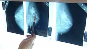
Another large study looking at screening mammograms for breast cancer has raised the issue of overdiagnosis and overtreatment once again. The purpose of mammography screening is to find cancer when it is small and so prevent cancer from growing and becoming advanced cancer. However, the researchers did not find this - there was a major increase in finding small cancers (the kind that may grow so slowly as to never cause any problems or that may even regress), but the rate of advanced cancers stayed the same.

The problem of overdiagnosis (finding small tumors that may never cause problems) and overtreatment (treating unnecessarily), which is leading to medical experts "rethinking cancer screening" is a major shift in how cancer screening is being viewed for a number of cancers. This is because studies show that overall death rates are basically the same in screened vs non-screened persons for mammography, colon, prostate, and lung cancer screening (see post). The view of how cancer grows and spreads may have to be reexamined and changed. One possibility suggested by Dr. H. Gilbert Welch is that aggressive cancer is already "a systemic disease by the time it's detectable" (Oct. 28, 2015 post).

The following excerpts are from the thoughtful review of the study in Health News Review: Overdiagnosis of ductal carcinoma in situ: ‘the pathology equivalent of racial profiling’

Danish researchers are providing new evidence that many breast cancers found via screening mammograms don’t need to be treated. Women with these non-threatening tumors are said to be “overdiagnosed” with breast cancerOverdiagnosis occurs when breast screening such as mammography detects small, slow-growing cancers that may never cause the patient any trouble. Yet, women diagnosed with such tumors are exposed to very real harms–possible surgery, chemotherapy, radiation, and living life as a “cancer patient.”

How much overdiagnosis are we talking about? If you don’t include cases of ductal carcinoma in situ (DCIS) in the tallies, anywhere from 14.7% to 38.6% of breast cancers found via screening represent overdiagnosis, the study authors found. The rate ranges from 24.4% to as high as 48.3% when DCIS is included.

DCIS is a collection of abnormal cells inside a milk duct that may–but usually doesn’t–break out to become invasive and potentially lethal cancer. About 60,000 women are told they have DCIS each year in the United States. Some experts estimate that up to 80% of women with DCIS found via screening may not need any treatment at all–and instead should just keep an eye on things. Obviously, women need to be fully and accurately informed about the benefits and risks — including the risk of overdiagnosis — before embarking on any decision to get screened for breast cancer or choosing a course of action following a diagnosis.

Otis Brawley, MD, Chief Medical Officer for the American Cancer Society, says it’s been difficult for modern medicine to wrap its brain around the concept of overdiagnosis. The natural inclination is to assume that cancerous-looking cells “will grow, spread, and eventually kill,” he writes in an editorial accompanying the Danish study. “However, some of these lesions may be genomically predetermined to grow no further and may even regress. In many respects, considering all small breast lesions to be deadly and aggressive types of cancer is the pathologic equivalent of racial profiling.

Excerpts from the original study from the Annals of Internal Medicine: Breast Cancer Screening in Denmark: A Cohort Study of Tumor Size and Overdiagnosis

Background: Effective breast cancer screening should detect early-stage cancer and prevent advanced disease. Objective: To assess the association between screening and the size of detected tumors and to estimate overdiagnosis (detection of tumors that would not become clinically relevant).... Setting: Denmark from 1980 to 2010. Participants: Women aged 35 to 84 years. Intervention: Screening programs offering biennial mammography for women aged 50 to 69 years beginning in different regions at different times.

Conclusion: Breast cancer screening was not associated with a reduction in the incidence of advanced cancer. It is likely that 1 in every 3 invasive tumors and cases of DCIS (ductal carcinoma in situ) diagnosed in women offered screening represent overdiagnosis (incidence increase of 48.3%).

Breast screening is associated with a substantial increase in the incidence of nonadvanced tumors and DCIS (ductal carcinoma in situ) in Denmark but not with a reduction in the incidence of advanced tumors, and the overdiagnosis rate is substantial. These findings support that screening has not accomplished the promise of a reduction in invasive therapy or disease-specific mortality.

I saw mention of this study in a number of places - that low vitamin D levels are linked to chronic headaches. A little too soon to know if that is really true -  the researchers in this study looked at the blood vitamin D levels of 2601 men just one time, and did not give vitamin D supplements to the men to see if this changed the frequency of migraine headaches. The researchers themselves pointed out that other studies looking at this same issue have had mixed results. And they themselves pointed out that low blood levels of vitamin D (serum 25(OH)D concentration) was associated with a markedly higher risk of frequent headache in men. Associated does not mean caused.

Yes, low vitamin D levels is linked to a number of health problems (see all vitamin D posts). But at this point I think that it's a case of "wait and see" to see if vitamin D levels have something to do with headache frequency. Perhaps other micronutrients are important, perhaps something else. Note that in the study they used below 50 nmol as the measure for low vitamin D levels. In the USA, that translates into below 20 ng/ml, which everyone agrees is too low (a deficiency). The best source of vitamin D is sunlight - which is why it's called the "sunshine vitamin". From Science Daily:

Vitamin D deficiency increases risk of chronic headache

Vitamin D deficiency may increase the risk of chronic headache, according to a new study from the University of Eastern Finland....The Kuopio Ischaemic Heart Disease Risk Factor Study, KIHD, analysed the serum vitamin D levels and occurrence of headache in approximately 2,600 men aged between 42 and 60 years in 1984-1989. In 68% of these men, the serum vitamin D level was below 50 nmol/l, which is generally considered the threshold for vitamin D deficiency. Chronic headache occurring at least on a weekly basis was reported by 250 men, and men reporting chronic headache had lower serum vitamin D levels than others.

When the study population was divided into four groups based on their serum vitamin D levels, the group with the lowest levels had over a twofold risk of chronic headache in comparison to the group with the highest levels. Chronic headache was also more frequently reported by men who were examined outside the summer months of June through September. Thanks to UVB radiation from the sun, the average serum vitamin D levels are higher during the summer months.

The study adds to the accumulating body of evidence linking a low intake of vitamin D to an increased risk of chronic diseases. Low vitamin D levels have been associated with the risk of headache also by some earlier, mainly considerably smaller studies.

Another large study has found negative health effects from living close to high-traffic roadways - this time a higher risk of dementia. The closer to the heavy traffic road, the higher the risk - with the highest risk in people living less than 50 meters (164 feet) from a high-traffic roadway, especially in major urban cities.

Other studies suggest that the air pollution from the high-traffic roadways is the problem - that the pollutants from vehicles (such as from the exhaust, and the wear of the tires) get into the body and brain and cause systematic inflammation. (see posts on air pollution and the brainTraffic-related air pollution includes ultrafine particles, nitrogen oxides, and particles. And the constant noise is stressful. However, this large Canadian study found no association with multiple sclerosis or Parkinson's disease and living close to heavy traffic roadways. (See all posts on air pollution and the brain.) From Science Daily:

Living near major traffic linked to higher risk of dementia

People who live close to high-traffic roadways face a higher risk of developing dementia than those who live further away, new research from Public Health Ontario (PHO) and the Institute for Clinical Evaluative Sciences (ICES) has found. Led by PHO and ICES scientists, the study found that people who lived within 50 metres of high-traffic roads had a seven per cent higher likelihood of developing dementia compared to those who lived more than 300 meters away from busy roads.

Published in The Lancet, the researchers examined records of more than 6.5 million Ontario residents aged 20-85 to investigate the correlation between living close to major roads and dementia, Parkinson's disease and multiple sclerosis....The findings indicate that living close to major roads increased the risk of developing dementia, but not Parkinson's disease or multiple sclerosis, two other major neurological disorders.

"Our study is the first in Canada to suggest that pollutants from heavy, day-to-day traffic are linked to dementia. We know from previous research that air pollutants can get into the blood stream and lead to inflammation, which is linked with cardiovascular disease and possibly other conditions such as diabetes. This study suggests air pollutants that can get into the brain via the blood stream can lead to neurological problems," says Dr. Ray Copes, chief of environmental and occupational health at PHO and an author on the paper.

People who lived within 50 metres of high-traffic roads had a seven per cent higher likelihood of dementia than those who lived more 300 meters away from busy roads. - The increase in the risk of developing dementia went down to four per cent if people lived 50-100 metres from major traffic, and to two per cent if they lived within 101-200 metres. At over 200 metres, there was no elevated risk of dementia. -There was no correlation between major traffic proximity and Parkinson's disease or multiple sclerosis. [Original study]

 Image result for breathing mouth What if you could detect cancer by a gadget that can sniff for cancer or some other disease? The theory that disease is detectable from exhaled breath dates back to about 400 BC when the Greek physician Hippocrates used to smell his patients' breath to find out what ailed them. More recently a number of individual diseases, such as diabetes, have been detected with "breath analysis".

But Israeli scientist Hossam Haick and others wanted to invent a medical diagnostic tool (an "electronic nose") that could detect multiple diseases, including cancer, by "sniffing" a patient’s exhaled breath. They recently published a study suggesting that such a device that they developed can work for 17 diseases (including 8 types of cancer, Crohn's disease, ulcerative colitis, irritable bowel syndrome, Parkinson's disease, multiple sclerosis, pre-eclampsia in pregnant women, and chronic kidney failure). The results showed that each of these diseases has its own unique "breathprint", and which overall could be detected with 86% accuracy using the device.

The study used breath samples collected from 1,404 people from 2011 to 2014 in 5 countries (Israel, the United States, Latvia, China, and France). The test subjects were either known to be healthy or to be suffering from one of the 17 diseases. Please note that this diagnostic tool is currently being further researched and developed. It is not available yet.

From Science Daily: Nanoarray sniffs out and distinguishes multiple diseases

Before modern medical lab techniques became available, doctors diagnosed some diseases by smelling a patient's breath. Scientists have been working for years to develop analytical instruments that can mimic this sniff-and-diagnose ability. Now, researchers report in the journal ACS Nano that they have identified a unique "breathprint" for each disease. Using this information, they have designed a device that screens breath samples to classify and diagnose several types of diseases.

Exhaled breath contains nitrogen, carbon dioxide and oxygen, as well as a small amount of more than 100 other volatile chemical components. The relative amounts of these substances vary depending on the state of a person's health. As far back as around 400 B.C., Hippocrates told his students to "smell your patients' breath" to search for clues of diseases such as diabetes (which creates a sweet smell). In more recent times, several teams of scientists have developed experimental breath analyzers, but most of these instruments focus on a single type of disease, such as cancer. In their own work, Hossam Haick and a team of collaborators in 14 clinical departments worldwide wanted to create a breathalyzer that could distinguish among multiple diseases.

The researchers developed an array of nanoscale sensors to detect the individual components in thousands of breath samples from patients who were either healthy or had one of 17 different diseases, such as kidney cancer or Parkinson's disease. By analyzing the results with artificial intelligence techniques, the team could use the array to classify and diagnose the conditions. The team used mass spectrometry to identify the breath components associated with the diseases. They found that each disease produces a unique volatile chemical breathprint, based on differing amounts of 13 components. They also showed that the presence of one disease would not prevent the detection of others -- a prerequisite for developing a practical device to screen and diagnose various diseases in a noninvasive, inexpensive and portable manner.

I've been asked whether vegan diets are safe during pregnancy. And I've always said that I don't know, but that avoiding all meat, fish, eggs, and dairy concerns me. Vegan diets are diets without meat, fish, dairy, honey, and eggs (no animal derived food), but while vegetarian diets also avoid meat, they do include eggs, honey, milk, and dairy products. Thus it is very important that anyone following a vegan diet plan meals carefully to get all the necessary nutrients. For example, soda and french fries are vegan, but are not good nutritionally.

There are a number of nutrients that probably need to be supplemented in vegan diets during pregnancy, especially B-12, iron, zinc, iodine (can get by using iodized salt), and omega-3s, and that's not even discussing micronutrients. So it was good to see this article by Dr. Drew Ramsey (Columbia University, NY) raising those same concerns about nutrient deficiencies during pregnancy, and pointing out that very few studies have looked at vegetarian and vegan diets in pregnancy - that the evidence is "scant". This topic is so controversial that as of today there were 85 comments by health professionals after the following Medscape article - both strongly pro and con, and studies given that show that vegans tend to have lower levels of a number of nutrients measured (zinc, iron, omega-3s, etc).

There are also reports of babies exclusively breast-fed by vegan mothers (here and here) who did not take vitamin supplements that developed nutritional deficiencies (especially B-12, vitamin K, and vitamin D). This is because what a mother eats is what the baby gets during pregnancy and breast-feeding.

On the other hand, the Academy of Nutrition and Dietetics came out with a position statement in 2016 stating that "appropriately planned vegetarian, including vegan, diets are healthful, nutritionally adequate, and may provide health benefits for the prevention and treatment of certain diseases. These diets are appropriate for all stages of the life cycle, including pregnancy, lactation, infancy, childhood, adolescence, older adulthood, and for athletes". But that article (in the Journal of Academy of Nutrition and Dietetics) then goes on in depth about several nutrients that can be lower or deficient in vegan diets unless supplements are given (B-12, iodine, etc). From Medscape;

Are Vegan and Vegetarian Diets Safe During Pregnancy?

Are vegan and vegetarian diets safe during pregnancy?....Vegan and vegetarian diets are plant-based diets....Plants, in general, are a great choice, especially colorful plants, because they tend to be more nutrient-dense. Plant-based diets have been linked to a number of health benefits, such as lower body mass index and lower rates of obesity and diabetes, as well as conferring some benefits. Certainly, vegan and vegetarian diets have higher amounts of certain nutrients like magnesium, folate, and fiber, all of which are generally consumed in very low quantities in Western diets.

Focusing on pregnancy, there are two important papers to note. The first, which got me very interested in this issue, was a series of pooled case reports by Drs Dror and Allen in 2008.[1] They looked at 30 cases of severe vitamin B12 deficiency during pregnancy in vegan women with pernicious anemia. Among the 30 vegan women who had B12 deficiency during pregnancy, about 60% of their offspring had severe developmental delays and 37% had cerebral atrophy.... The clinical importance of this is to partner with our patients who are eating plant-based diets and ensure that they have adequate levels of vitamin B12, as well as iron, zinc, and long-chain omega-3 fats during pregnancy.

The second article to take a peek at is from the British Journal of Obstetrics and Gynaecology in 2015.[2] This was a systematic review that looked at all of the literature after screening out the papers. The authors found about 13 papers focusing on maternal and infant outcomes, and about nine of those looked at nutrient deficiency. The main headline of this review is that there are no randomized clinical trials of vegan or vegetarian diets in pregnancy. It is very hard to make a clear clinical recommendation. The epidemiologic data were heterogeneous. As the researchers called it, it was "scant." That is certainly true. Overall, there were no clear associations with bad outcomes. There was some increased risk for hypospadias in one of the larger trials that looked at vegan and vegetarian diets during pregnancy.

The main concern is that vegan and vegetarian diets put patients at risk for a number of nutrient deficiencies—vitamin B12 in vegan diets (and even for vegetarians), iron, zinc, and the long-chain omega-3 fats. Just a quick moment on the long-chain omega-3 fats: Dietarily, these only come from fatty fish and seafood. They are bioconcentrated. They start in algae, but they get bioconcentrated in our food supply via seafood. It is very hard to get those during pregnancy. Certainly, a lot of healthy babies have been born to vegetarian and vegan women. You can supplement, but given the benefits we see with omega-3 fats in patients with things like mood disorders or a history of psychotic disorders, I think it is important to consider the long-chain omega-3 fats. If someone is not going to get those in their diet but has a history, risk, or propensity toward mood disorders, think about supplementation. 

Excerpts from the review study (mentioned above) in BJOG: Vegan-vegetarian diets in pregnancy: danger or panacea? A systematic narrative review.

To review the literature on vegan-vegetarian diets and pregnancy outcomes [in healthy pregnant women]....None of the studies reported an increase in severe adverse outcomes or in major malformations, except one report of increased hypospadias in infants of vegetarian mothers. Five studies reported vegetarian mothers had lower birthweight babies, yet two studies reported higher birthweights. The duration of pregnancy was available in six studies and was similar between vegan-vegetarians and omnivores. The nine heterogeneous studies on microelements and vitamins suggest vegan-vegetarian women may be at risk of vitamin B12 and iron deficiencies.

CONCLUSIONS: The evidence on vegan-vegetarian diets in pregnancy is heterogeneous and scant. The lack of randomised studies prevents us from distinguishing the effects of diet from confounding factors. Within these limits, vegan-vegetarian diets may be considered safe in pregnancy, provided that attention is paid to vitamin and trace element requirements.

1

 Two recent reviews of studies looked at erectile dysfunction from different angles. The first review found that regular exercise or physical activity improves erectile function  (especially if the person has heart disease or has had their prostate removed) - with improvements similar to what people see with a medication. Other studies have found similar results (see here and here). The other review found that having chronic periodontitis (gum disease) is linked to a higher incidence of erectile dysfunction, with one study finding that treating the periodontitis improves erectile dysfunction symptoms. Bottom line: get regular physical activity and exercise, and get regular dental care (and treat any periodontitis).

From Medscape: Erectile Dysfunction May Improve With Exercise

Men who have difficulty maintaining erections may benefit from exercise or physical activity, according to a new analysis. A growing body of evidence has suggested physical activity could improve erectile function, but the authors of the new report say that until now, no one had looked at all the studies together....The prevalence of erectile dysfunction ranges from 8 percent among men in their 20s and 30s, up to 37 percent among men in their early 70s.

For the new study, the researchers searched for randomized controlled trials testing exercise and physical activity as a therapy for erectile dysfunction. They found seven studies published between 2004 and 2013 that included a total of 505 men, who were tracked for anywhere from eight weeks to two years. Average ages ranged from 43 to 69 years. Altogether, 292 men were randomly assigned to complete aerobic exercises, pelvic floor muscle exercises or a combination of exercises. The other 213 men were not told to exercise.

Erectile function was measured using the International Index of Erectile Function. Scores range from 5 to 25; men with no erectile dysfunction have scores of 22 to 25, and those with the most severe dysfunction score between 5 and 7. Overall, men who exercised had their scores increase by an average of 3.85 points, compared to men who did not exercise. Exercises specific to pelvic floor muscles didn't seem to yield a benefit. Among men with an increased cardiovascular risk, coronary heart disease or prostate removal, however, any type of exercise led to improved erectile function scores.

A take-home message from this analysis is that exercise should have a role in the treatment of erectile dysfunction, said Dr. Landon Trost, who is head of andrology and male infertility at the Mayo Clinic in Rochester, Minnesota....He told Reuters Health that the average increase in erectile function score would likely be similar to what people see with a medication.

From Medscape: Periodontitis Tied to Risk of Erectile Dysfunction

Erectile dysfunction is more common in men with chronic gum disease, according to a new review of existing studies. Periodontitis has been tied to a higher risk of cardiovascular disease and general inflammation, which in turn have been tied stroke and atherosclerosis, which are associated with erectile dysfunction (ED). 

The reviewers analyzed data from five studies published between 2009 and 2014, including one randomized controlled trial. In total, the studies covered 213,000 participants aged 20 to 80. Each study found erectile dysfunction was more common among men being treated for chronic periodontitis, particularly for those younger than 40 and older than 59. After accounting for diabetes, which can influence both gum disease and sexual function, erectile dysfunction was 2.28 times more common for men with periodontitis than for men without it, according to the report in the International Journal of Impotence Research....One study in 2013 found that treating periodontitis improves erectile dysfunction symptoms.

The new review did have limitations, including the fact that erectile dysfunction and chronic periodontitis are caused by similar risk factors, such as aging, smoking, diabetes mellitus and coronary artery disease. While some studies did account for diabetes, most did not account for smoking or alcohol consumption, which can also affect oral health and sexual function, the authors write. Even after an exhaustive search, the evidence linking periodontitis and ED is limited, they write. 

Tobacco use is a leading cause of cancer and early death in the U.S. and throughout the world. According to a new study looking at people 70 years old and older, the good news is that quitting smoking at any time in life (even as late as the 60s) is better for immediate health and also reduces the risk of death.

The study compared people who had never smoked to people currently smoking - and found that in the 6 year follow up period current smokers were more than three times more likely to die than people who had never smoked. Furthermore, quitting smoking at any age was associated with a lower risk of death. Former smokers who quit smoking earlier in life received the largest benefit from quitting smoking. But even people who quit during their 60s were at substantially decreased risk of death (when compared to people who continued to smoke). Bottom line: It's never too late to quit smoking. But best is to never even start smoking. From MedicalXpress:

Quitting smoking at any age reduces the risk of death after 70

Tobacco use continues to be a major cause of cancer and premature death. Most studies of cigarette smoking and mortality have focused on middle-aged populations, with fewer studies examining the impact of tobacco cessation on disease and mortality risk among the elderly. A new study published in the American Journal of Preventive Medicine, found that people aged 70 or older currently smoking were more than three times more likely to die than never-smokers, while former smokers were less likely to die the sooner they quit.

Investigators reviewed data for more than 160,000 individuals aged 70 and over who participated in the NIH-AARP Diet and Health Study. They completed a questionnaire in 2004-2005 detailing their smoking use, and reported deaths were tracked until the end of 2011....For this study, participants still smoking in their 70s were identified as current smokers, and former smokers were classified by the decade of life when quitting. At the beginning of the study (2004-2005), the median age of participants was 75 years. Almost 56% were former smokers and 6% were current smokers. Males were less likely (31% vs 48% of females) to be never-smokers. 

During an average follow-up of 6.4 years, almost 16% of the participants died. While 12.1% of the never smokers died, 16.2%, 19.7%, 23.9%, and 27.9% of former smokers who quit between ages 30-39, 40-49, 50-59, and 60-69 years died, respectively. Current smokers fared the worst, with 33.1% dying. Mortality rates for women were lower than men at each level of smoking use.

"These data show that age at smoking initiation and cessation, both key components of smoking duration, are important predictors of mortality in U.S. adults aged 70 years and older," commented Dr. Nash. "In the NIH-AARP study population, younger age at initiation was associated with increased risk of mortality, highlighting the importance of youth and early-adult smoking on lifetime mortality risk, even among people who live to age 70 years. In addition, former smokers were at substantially reduced risk of mortality after age 70 years relative to current smokers, even those who quit in their 60s. These findings show that smoking cessation should be emphasized to all smokers, regardless of age."

A recent article discussed the large assortment of medications (both prescription and non-prescription) that are linked to liver injury, commonly known as "Drug-induced liver injury" (DILI). While it occurs rarely (fewer than 10 in 10,000 persons who take the drug in question), many medications can result in liver injury - especially if taken in too large doses and for too long. The scary part is that 46% of people with acute liver failure in the U.S have the liver damage from acetaminophen. Acetaminophen is the main cause of drug induced liver injury and liver failure in the U.S. Acetaminophen is found not just in Tylenol, but in many non-prescription drugs - thus it is easy to take too large a dose. The liver helps remove toxins - thus we need to take good care of it. From Science Daily:

Acetaminophen, supplements and other medications may trigger drug-induced liver injury

More than 1,000 medications, with acetaminophen being the most common, have been associated with drug-induced liver injury (DILI). Diagnosis can be challenging due to the multitude of contributing factors, and timely recognition and clinical response may mean the difference between recovery and acute liver failure or even death. DILI affects an estimated fewer than 10 people in every 10,000 exposed persons. The condition is dose-dependent or an adverse reaction to a medication, dietary supplement or other substance. 

"The liver helps remove toxins, which makes it especially vulnerable to injury from either short-term intake above recommended levels or long-term usage that allows toxins to build up," Collins-Yoder said. "Recognizing the clinical signs and symptoms is crucial to prompt treatment and effective patient care." Depending on the contributing factors and the level of damage to the liver, patients with mild and moderate signs and symptoms may recover normal liver function after the triggering substance is identified and use is discontinued. Other patients may experience more severe damage, progressing to acute liver failure.

About 46 percent of persons with acute liver failure in the United States have liver damage associated with acetaminophen, making it the most common cause of DILI. Since acetaminophen is often an ingredient in over-the-counter and prescription pain medications, patients may take higher doses than needed. A more infrequent type of DILI is triggered by an adverse reaction to prescription medications, herbal dietary supplements or other substances, including: nonsteroidal anti-inflammatory drugs (NSAIDs), including ibuprofen, naproxen and others; antibiotics and antiviral agencies, such as amoxicillin-clavulanate, sulfamethoxazole-trimethoprim and nitrofurantoin; antileptic agents, such as volproic acid and carbamazepine; statins; novel anticoagulants; proton pump inhibitors; methotrexate; azathioprine; sulfasalazine; herbal and dietary supplements.

Study after study finds negative health effects from air pollution, such as heart disease (here, here, and here). Now two more studies found that living in areas with high air pollution is linked to a higher stroke risk. One study (done in Japan) found an increase of ischemic stroke on the same day as exposure to high levels of air pollution, while the other (done in London, UK) found a higher risk of death after stroke (especially ischemic strokes) in patients who live in areas of high air pollution. This was especially pronounced with exposure to smaller or fine particulate matter (PM2.5)- which is found in high quantities in vehicle exhaust fumes.

It is thought that the fine particles in the air (PM2.5) contribute to the development of potentially fatal diseases various ways - by causing chronic inflammation, and also because they slip past the body's defenses and can be absorbed deep into the lungs and bloodstream. They are not sneezed or coughed out the way larger natural particles, like airborne soil and sand, are removed from the body's airways. What can be done? Other studies have found that when air pollution is reduced, than the risk of death is reduced. So yes, pollution controls on vehicles such as trucks and buses are good. And just think how much air pollution will be reduced when electric vehicles replace current gas powered cars and trucks. From Medscape:

Air Pollution Linked to Higher Stroke Mortality

More evidence showing that living in areas with high levels of air pollution is linked to a higher stroke risk has come from two new studies. Both studies are published online in the journal Stroke. The first, from the United Kingdom, shows a higher risk for death after a stroke in patients who live in areas of high air pollution, and the other, a Japanese study, suggests a higher risk for a new stroke the same day as exposure to high levels of air pollution.

"We have shown a significantly increased risk of death after stroke in patients who had long-term exposure to high levels air pollution before their stroke occurred," senior author of the UK study, Charles Wolfe, MD.... "This was particularly pronounced for high exposure to smaller particulate matter — particles below 2.5 μm in diameter (PM2.5) — which are found in high quantities in exhaust fumes." For the study, Professor Wolfe and colleagues analyzed data from the South London Stroke Register, a population-based register covering an urban, multiethnic population.....Results showed an increased risk for death up to 5 years after stroke in patients living in areas of high air pollution.

"While this study adds to the evidence linking air pollution to cardiovascular disease, it cannot prove causality as it has an observational design," Professor Wolfe commented...."So it is difficult to say for certain that it is the air pollution that is responsible but there are many studies now that have shown similar associations," he said. He noted that the smaller particles (PM2.5) were associated with a worse effect on mortality and this correlated with biological studies that have shown a greater inflammatory effect of small particulate matter vs larger particulate matter on the vessel wall. "Our study suggests that people who have previously had an ischemic stroke, but not a hemorrhagic stroke, may be more vulnerable and at a higher risk of death to chronic, long-term exposure of PM," they conclude.

The Japanese study, by lead author Ryu Matsuo, MD, PhD, Department of Health Care Administration and Management Graduate School of Medical Sciences, Kyushu University, Fukuoka, Japan, and colleagues, looked at acute exposure to air pollution and shows a small increase in the risk of having a stroke within a day of high exposure to pollution. For the study, the researchers analyzed data on 6885 ischemic stroke patients from a multicenter hospital-based stroke registry in Japan who were previously independent and hospitalized within 24 hours of stroke onset.

Professor Wolfe said his group have conducted a similar study looking at exposure of air pollution in the year before stroke, which showed a 23% increase in stroke risk in those exposed to higher levels of PM2.5. 

An important study was recently published that documents that when pregnant women are exposed to potentially harmful chemicals in the environment around them (environmental pollutants), many of these chemicals also get transferred to the fetus. Many of the 59 chemicals looked for were detected in the newborn babies' umbilical cords (thus the chemicals had crossed the placenta to the fetus during the pregnancy). Some chemicals were measured in higher levels in the umbilical cord than in the mother (as measured in her blood), while only cadmium appeared in mothers, but not in the umbilical cord (thus there was some protection for the fetus from that particular chemical).

This study did not look at the effects on the fetus and newborn from exposure to all these toxic chemicals - it just found that out of 59 chemicals tested for, the median number was 25 in maternal blood and 17 in umbilical cord blood. Eight of the 59 chemicals analyzed were detected in more than 90 percent of both the maternal and cord blood samples - and these included lead and mercury.

The researchers pointed out that no human studies have examined the developmental and reproductive health effects on the fetus and baby from being exposed to multiple chemicals simultaneously during pregnancy - which of course can have bigger risks and negative effects than being exposed to only one chemical at a time. Other studies have already shown that there are negative health effects (adverse neurodevelopmental effects) from a number of these chemicals, such as PCBs, PBDEs, Pb (lead) and Hg (mercury). Bottom line: No one really knows what these mixtures of chemicals do to the developing baby, but it is known that these chemicals have potential health risks. From Futurity:

Mom’s exposure to toxic chemicals shows up in newborn

Low-income and Latina pregnant women in a recent study had widespread exposure to environmental pollutants. In addition, many of the toxins showed up at even higher levels in their newborns. The study is the first in the United States to measure exposure to 59 toxic chemicals in pregnant women and their newborns.

“Pregnant women in the US are exposed to many harmful industrial chemicals that have been linked to premature birth, low birth weight, and birth defects, but estimates of how efficiently pollutants are transferred from mother to fetus have varied widely,” says Tracey Woodruff, professor of obstetrics, gynecology, and reproductive sciences and the Philip R. Lee Institute for Health Policy Studies at the University of California, San Francisco. “Our findings have found that many chemicals do indeed accumulate in the fetal environment and are absorbed at greater levels by fetuses than by the pregnant women themselves. This may have significant consequences for the growing fetus, since many of these chemicals are known to affect development.”

Researchers measured polychlorinated biphenyls (PCBs), organochlorine pesticides (OCPs), polybrominated diphenyl ethers (PBDEs), perfluorinated compounds (PFCs), mercury, and lead, among other chemicals. These industrial pollutants are common in the environment, and in previous studies many have been detected in greater than 99 percent of US pregnant women, according to National Health and Nutrition Examination Survey (NHANES) data.

“Contrary to previous research, we found evidence that several PCBs and OCPs were often higher in umbilical cord samples than in maternal blood samples,” says Rachel Morello-Frosch,  professor of environmental science, policy and management at the University of California, Berkeley. The study also found that concentrations of mercury and certain PBDEs were often higher in umbilical cord samples than in maternal samples, and for most PFCs and lead, cord blood concentrations were generally equal to or lower than maternal concentrations, which is consistent with previous research.

Almost 80 percent of the chemicals detected in maternal blood samples were also detected in the umbilical cord blood samples, indicating that they passed through the placenta and entered the fetal environment, where they can pose a health risk to the developing baby. For those chemicals detected in at least 20 paired maternal and umbilical cord samples, 77 percent had significant correlations between maternal and umbilical cord concentrations.

The women in the study were participating in the Chemicals in Our Bodies Study, also referred to as the Maternal and Infant Environmental Exposure Project. Of the women participating in the current study, 95 percent had a combined annual household income of less than $40,000, two-thirds were Latina, and a third were born in Mexico, where they may have had less exposure to environmental toxics like the PBDEs found in flame retardants that have been widely used in the US. This demographic is often not well-represented in larger biomonitoring studies, such as NHANES, that form the basis of most of what is known about pregnant women’s exposure to environmental toxics nationally.

The study also provides a first indication of how several different classes of environmental chemicals found in a pregnant woman’s blood are also present in the newborn....From 2010 to 2011, researchers collected maternal blood samples from 77 pregnant women at Zuckerberg San Francisco General. Once they delivered their babies, researchers collected umbilical cord blood samples from 65 of these women. Of those samples tested for all 59 chemicals, the median number was 25 in maternal blood and 17 in umbilical cord blood. Eight of the 59 chemicals analyzed were detected in more than 90 percent of both the maternal and cord blood samples.