Skip to content

A thought-provoking article by Heiman and Greenway was just published in the journal Molecular Metabolism making the case that changes in farming practices over the last 50 years have resulted in decreased agricultural diversity which, in turn, has resulted in decreased dietary diversity, and that the reduction in dietary diversity has changed and decreased the richness of the human gut microbiota (microbes living in the gut). And meanwhile, during the past 50 years, the rates of obesity, type 2 diabetes, and inflammatory bowel diseases sharply increased - and in each of these conditions there is a reduction of the gut microbial diversity. Similar views have also been stated by others in the field of microbiology.

The thinking is that the more diverse the diet, the more diverse the gut microbiome (and healthier), and the more it can adapt to disturbances. Heiman and Greenway state: "Unfortunately, dietary diversity has been lost during the past 50 years because of economic pressures for greater food production to support a growing world population.... Of the 250,000 to 300,000 known edible plant species, humans use only 150 to 200...Today, 75 percent of the world's food is generated from only 12 plants and five animal species."

Also, agricultural practices of using antibiotics as growth promoters for poultry, swine, and cattle further harm the human gut microbiome when the meat is ingested by humans, and pesticide residues on crops ingested by humans may have gut microbiome effects. Even emulsifiers, used in processed foods, reduce microbial richness. Every time a person goes on a certain diet (vegan, Paleo, etc) or makes dietary choices in which some foods are eliminated, it makes it easier for some microbial species, and gives them a competitive advantage over other gut microbes. From Science Daily:

Reduction in dietary diversity impacts richness of human gut microbiota

Changes in farming practices over the last 50 years have resulted in decreased agro-diversity which, in turn, has resulted in decreased dietary diversity. The significant impact of this change in dietary richness on human health is an emerging topic for discussion

Heiman and Greenway describe how the reduction in dietary diversity has changed the richness of human gut microbiota, the community of microorganisms living in the gut. The researchers point out that healthy individuals have diverse gut microbiota and many of the common pathologies of the 21st century, including type 2 diabetes, obesity and inflammatory bowel disease, are associated with reduced microbiotic richness.

Gut microbiota function as an endocrine organ, metabolizing specific nutrients from the diet and producing specific substances that act as metabolic signals in the host. It follows then that highly specialized diets will change the landscape of the gut microbiome over time. In fact, it takes only a few days of changing diet to alter the microbiotic makeup of the human gut. And if the dietary change involves elimination of one or more macronutrients (think Atkins or Paleo or vegan), humans are essentially selecting for some microbiotic species over others.

The importance of microbiota diversity cannot be overstated. They produce an abundance of important molecules for the host and with increased variation comes increased adaptability and an increased range of physiological responses. "The greater the repertoire of signals, the more likely is the ability to maintain homeostasis when dietary intake is perturbed," explain Heiman and Greenway. "Furthermore, because each particular macronutrient has the potential to be metabolized by microbiota into unique metabolic signals, the greater the variety in signals, the greater the variety of responses possible."

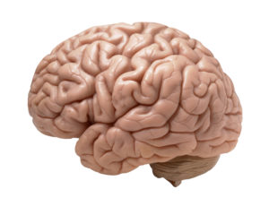
 Two articles about the link between Alzheimer's disease (AD) and microbes this past week: a study linking periodontal disease and Alzheimer's, and the other a journal editorial (written by an international team of 31 researchers) suggesting that we need to more closely look at the role of microbes in Alzheimer's disease, especially herpes virus, chlamydia and spirochaete bacteria.

This team is suggesting an "infectious cause" for Alzheimer's, an example being the reactivation of herpes simplex virus type 1 (HSV1) in the person. The researchers state that "regarding HSV1, about 100 publications by many groups indicate directly or indirectly that this virus is a major factor in the disease".  The team also mentions the possibility of fungi infection in some cases (see my November 6, 2015 post about a study finding fungal involvement). Both articles mention that treatment of the diseases with some form of antimicrobials or antivirals could possibly treat Alzheimer's disease, and that trails now need to be done.

From Science Daily: Link between gum disease and cognitive decline in Alzheimer’s

A new study has found a link between gum disease and greater rates of cognitive decline in people with early stages of Alzheimer's Disease. Periodontitis or gum disease is common in older people and may become more common in Alzheimer's disease because of a reduced ability to take care of oral hygiene as the disease progresses. Higher levels of antibodies to periodontal bacteria are associated with an increase in levels of inflammatory molecules elsewhere in the body, which in turn has been linked to greater rates of cognitive decline in Alzheimer's disease in previous studies.

The presence of gum disease at baseline was associated with a six-fold increase in the rate of cognitive decline in participants over the six-month follow-up period of the study. Periodontitis at baseline was also associated with a relative increase in the pro-inflammatory state over the six-month follow-up period. The authors conclude that gum disease is associated with an increase in cognitive decline in Alzheimer's Disease, possibly via mechanisms linked to the body's inflammatory response.....However, growing evidence from a number of studies links the body's inflammatory response to increased rates of cognitive decline, suggesting that it would be worth exploring whether the treatment of gum disease might also benefit the treatment of dementia and Alzheimer's Disease.

From Journal of Alzheimer's Disease: Microbes and Alzheimer’s Disease

We are researchers and clinicians working on Alzheimer’s disease (AD) or related topics, and we write to express our concern that one particular aspect of the disease has been neglected, even though treatment based on it might slow or arrest AD progression. We refer to the many studies, mainly on humans, implicating specific microbes in the elderly brain, notably herpes simplex virus type 1 (HSV1), Chlamydia pneumoniae, and several types of spirochaete, in the etiology of AD [1–4]. Fungal infection of AD brain [5, 6] has also been described, as well as abnormal microbiota in AD patient blood [7]. The first observations of HSV1 in AD brain were reported almost three decades ago [8]. The ever-increasing number of these studies (now about 100 on HSV1 alone) warrants re-evaluation of the infection and AD concept.

AD is associated with neuronal loss and progressive synaptic dysfunction, accompanied by the deposition of amyloid-β (Aβ) peptide, a cleavage product of the amyloid-β protein precursor (AβPP), and abnormal forms of tau protein, markers that have been used as diagnostic criteria for the disease [9, 10]. These constitute the hallmarks of AD, but whether they are causes of AD or consequences is unknown. We suggest that these are indicators of an infectious etiology. In the case of AD, it is often not realized that microbes can cause chronic as well as acute diseases; that some microbes can remain latent in the body with the potential for reactivation, the effects of which might occur years after initial infection; and that people can be infected but not necessarily affected, such that ‘controls’, even if infected, are asymptomatic [2].

Regarding HSV1, about 100 publications by many groups indicate directly or indirectly that this virus is a major factor in the disease....The only opposing reports, two not detecting HSV1 DNA in elderly brains and another not finding an HSV1–APOE association, were published over a decade ago [76–78]. However, despite all the supportive evidence, the topic is often dismissed as ‘controversial’. One recalls the widespread opposition initially to data showing that viruses cause some types of cancer, and that a bacterium causes stomach ulcers.

In summary, we propose that infectious agents, including HSV1, Chlamydia pneumonia, and spirochetes, reach the CNS and remain there in latent form. These agents can undergo reactivation in the brain during aging, as the immune system declines, and during different types of stress (which similarly reactivate HSV1 in the periphery). The consequent neuronal damage— caused by direct viral action and by virus-induced inflammation— occurs recurrently, leading to (or acting as a cofactor for) progressive synaptic dysfunction, neuronal loss, and ultimately AD.

AD causes great emotional and physical harm to sufferers and their carers, as well as having enormously damaging economic consequences. Given the failure of the 413 trials of other types of therapy for AD carried out in the period 2002–2012 [79], antiviral/antimicrobial treatment of AD patients, notably those who are APOE ɛ 4 carriers, could rectify the ‘no drug works’ impasse. We propose that further research on the role of infectious agents in AD causation, including prospective trials of antimicrobial therapy, is now justified.

Very exciting research IF it pans out - the idea of treating (some) cancers with probiotics (beneficial bacteria). This study was done on mice, and some mice started the probiotic mixture one week before they gave the mice the liver cancer, so...more limitations there. But the idea is so tantalizing and wonderful... And what was in the mixture of bacteria (called probiotic Prohep) that the mice ate that had beneficial results of shrinking liver tumors? The probiotic Prohep is composed of Lactobacillus rhamnosus GG (LGG), Escherichia coli Nissle 1917 (EcN), and heat inactivated VSL#3 (1:1:1).  VSL#3 contains: Streptococcus thermophilus, Bifidobacterium breve, Bifidobacterium longum, Bifidobacterium infantis, Lactobacillus acidophilus, Lactobacillus plantarum, Lactobacillus paracasei, and Lactobacillus delbrueckii. Note that Lactobacillus rhamnosus and some of the others are already found in many probiotic mixtures. From Medical Xpress:

Probiotics dramatically modulate liver cancer growth in mice

Medical research over the last decade has revealed the effects of the gut microbiome across a range of health markers including inflammation, immune response, metabolic function and weight....Previous studies have demonstrated the beneficial role of probiotics in reducing gastrointestinal inflammation and preventing colorectal cancer, but a new study published in the Proceedings of the National Academy of Sciences explored their immunomodulatory effects on extraintestinal tumors: specifically, in hepatocellular carcinoma (HCC)HCC is the most common type of liver cancer, and though it is relatively uncommon in the United States, it's the second-most deadly type of cancer worldwide and is particularly prevalent in regions with high rates of hepatitis. 

The researchers designed a study in a mouse model of HCC that quantified the immunological effects of a novel probiotic formulation called Prohep. They fed the mice Prohep for a week prior to tumor inoculation, and they observed a 40 percent reduction of tumor weight and size compared with control animals. Further, they established that the beneficial effects of the probiotics were closely related to the abundance of beneficial bacteria promoted by Prohep. These bacteria produce anti-inflammatory metabolites, which regulated pro-inflammatory immune cell populations via crosstalk between the gut and the liver tumor.

Among their findings, the researchers report that the probiotics reduced liver tumor growth by inhibiting angiogenesis, the process by which the body generates new blood vessels from existing ones, which is essential for tumor growth. They found significantly raised levels of hypoxic GLUT-1+, indicating that tumor reductions were due to hypoxia caused by reduced blood flow. Further, the tumors in the treated mice had 52 percent lower blood vessel area and 54 percent fewer vessel sprouts than the untreated mice.

They also determined that Prohep treatment down-regulated IL-17, a pro-inflammatory angiogenic factor. Because HCC is a highly vascularized tumor, the cancer is generally associated with high levels of IL-17 and an immune T-cell called T helper 17 (Th17), which is transported from the gut to HCC tumors via circulation. The researchers believe that reduced Th17 in tumor cells impedes the inflammation and angiogenesis and limits tumor growth. It's not surprising that they also found that probiotics increased the anti-inflammatory bacteria and metabolites present in the guts of treated mice. They conclude that Prohep intake has the capability of inhibiting tumor progression by modulating the gut microbiota.

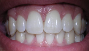
The finding that the oral bacteria Streptococcus mutans, which is found in 10% of the population, is linked with hemorrhagic strokes is big. S. mutans is found in tooth decay or cavities (dental caries). The researchers found a link with cnm-positive S. mutans with both intracerebral hemorrhage (ICH) and also with cerebral microbleeds.

Some risk factors for strokes have long since been known, such as high blood pressure and advanced age, but then there are those hemorrhagic strikes that don't seem to fit the norm, with no apparent risk factors. Well, apparently the presence of cnm-positive S. mutans is one. My understanding of what cnm-positive S. mutans means is S. mutans bacteria that carries the collagen-binding Cnm gene. This bacteria can be found in a person's saliva and in dental plaque, and swabs of both were taken for this study.

This study builds on other studies that find a link between the bacteria Streptococcus mutans and a number of systemic diseases, including bacteremia, infective endocarditis and hemorrhagic stroke. The researchers of this latest study suggest that infection with cnm-positive S. mutans causes constant inflammation (as shown by 2 inflammatory markers: CRP and fibrinogen), which then causes damage to blood vessels (endothelial damage) in the brain. Bottom line: take care of your teeth and gums.

From Science Daily: Oral bacteria linked to risk of stroke

In a study of patients entering the hospital for acute stroke, researchers have increased their understanding of an association between certain types of stroke and the presence of the oral bacteria (cnm-positive Streptococcus mutans).

In the single hospital study, researchers at the National Cerebral and Cardiovascular Center in Osaka, Japan, observed stroke patients to gain a better understanding of the relationship between hemorrhagic stroke and oral bacteria. Among the patients who experienced intracerebral hemorrhage (ICH), 26 percent were found to have a specific bacterium in their saliva, cnm-positive S. mutans. Among patients with other types of stroke, only 6 percent tested positive for the bacterium.

Strokes are characterized as either ischemic strokes, which involve a blockage of one or more blood vessels supplying the brain, or hemorrhagic strokes, in which blood vessels in the brain rupture, causing bleeding.

The researchers also evaluated MRIs of study subjects for the presence of cerebral microbleeds (CMB), small brain hemorrhages which may cause dementia and also often underlie ICH. They found that the number of CMBs was significantly higher in subjects with cnm-positive S. mutans than in those without. The authors hypothesize that the S. mutans bacteria may bind to blood vessels weakened by age and high blood pressure, causing arterial ruptures in the brain, leading to small or large hemorrhages.

"This study shows that oral health is important for brain health. People need to take care of their teeth because it is good for their brain and their heart as well as their teeth," Friedland said. "The study and related work in our labs have shown that oral bacteria are involved in several kinds of stroke, including brain hemorrhages and strokes that lead to dementia."

Multiple research studies have shown a close association between the presence of gum disease and heart disease, and a 2013 publication by Jan Potempa, Ph.D., D.Sc., of the UofL School of Dentistry, revealed how the bacterium responsible for gum disease worsens rheumatoid arthritisThe cnm-negative S. mutans bacteria is found in approximately 10 percent of the general population, Friedland says, and is known to cause dental cavities (tooth decay). Friedland also is researching the role of oral bacteria in other diseases affecting the brain.  http://www.nature.com/articles/srep20074

A recent study has examined the issue of whether the 10 to 1 ratio of bacteria to human cells, which is widely quoted, is actually correct. Weizmann Institute of Science researchers currently feel that based on scientific evidence (which of course will change over time) and making "educated estimates", the actual ratio is closer to 1:1 (but overall there still are more bacterial than human cells). They point out that the 10:1 ratio was originally a "back of the envelope" estimate dating back to 1972.

The researchers also point out that the ratio may vary over the course of each day - as a person defecates out huge amounts of bacteria with each bowel movement. However, this study - which is not the final word - is an educated guess about bacteria only. What about the viruses, the fungi, etc that also reside on and within us? We know much less about all the other microbes. I am disturbed that article after article, and headline after headline, equates microbes and bacteria. Microbes does not mean only bacteria.  From Science Daily:

Germs, humans and numbers: New estimate revises our microbiome numbers downwards

How many microbes inhabit our body on a regular basis? For the last few decades, the most commonly accepted estimate in the scientific world puts that number at around ten times as many bacterial as human cells. In research published in the journal Cell, a recalculation of that number by Weizmann Institute of Science researchers reveals that the average adult has just under 40 trillion bacterial cells and about 30 trillion human ones, making the ratio much closer to 1:1.

The rising importance of the microbiome in current scientific research led the Weizmann Institute's Prof. Ron Milo, Dr. Shai Fuchs and research student Ron Sender to revisit the common wisdom concerning the ratio of "personal" bacteria to human cells.

The original estimate that bacterial cells outnumber human cells in the body by ten to one was based on, among other things, the assumption that the average bacterium is about 1,000 times smaller than the average human cell. The problem with this estimate is that human cells vary widely in size, as do bacteria. For example, red blood cells are at least 100 times smaller than fat or muscle cells, and the microbes in the large intestine are about four times the size of the often-used "standard" bacterial cell volume. The Weizmann Institute scientists weighted their computations by the numbers of the different-sized human cells, as well as those of the various microbiome cells. 

Some excerpts from the original journal article from Cell: Are We Really Vastly Outnumbered? Revisiting the Ratio of Bacterial to Host Cells in Humans

The human microbiome has emerged as an area of utmost interest....One of the most fundamental and commonly cited figures in this growing field is the estimate that bacteria residing in the human body outnumber human cells by a factor of 10 or more (Figure 1A). This striking statement often serves as an entry point to the field. After all, if a human being is a cell population composed of at least 90% bacteria, it is only natural to expect a major role for them in human physiology.

Both the numerator (number of microbial cells) and the denominator (human cells) of this 10:1 ratio are based on crude assessments. Most sources cite the number of human cells as 1013 or 1014.....We performed a thorough review of the literature and found a long chain of citations originating from one “back of the envelope” estimate (Figure 1). This estimate, though illuminating, was never meant as the final word on the question.

Recently, the estimate of a 10:1 bacterial to human cell ratio (B/H) ratio has received criticism (Rosner, 2014). Therefore, an alternative value and an estimate of the uncertainty range are needed. Bacteria are found in many parts of the human body primarily on the external and internal surfaces, including the gastrointestinal tracts, skin, saliva, oral mucosa, and conjunctiva. The vast majority of commensal bacteria reside in the colon, with previous estimates of about 1014 bacteria (Savage, 1977), followed by the skin, which is estimated to harbor ∼1012 bacteriaBerg, 1996). Less than 1012 bacteria populate the rest of the body.....Almost all recent papers in the field of gut microbiota directly or indirectly rely on a single paper (Savage, 1977) discussing the overall number of bacteria in the gut. Interestingly, review of the original Savage 1977 paper demonstrates that it actually cites another paper for the estimate (Luckey, 1972)....The estimate, performed by Luckey in 1972, is an illuminating example of a back-of-the-envelope estimate, which was elegantly performed, yet was probably never meant to serve as the cornerstone reference number to be cited decades later.

Updating the ratio of bacteria to human cells from 10:1 or 100:1 to closer to 1:1 does not take away from the biological importance of the microbiota. ...Although we still appear to be outnumbered, we now know more reliably to what degree and can quantify our uncertainty about the ratios and absolute numbers. The B/H ratio is actually close enough to one, so that each defecation event, which excretes about 1/3 of the colonic bacterial content, may flip the ratio to favor human cells over bacteria. This anecdote serves to highlight that some variation in the ratio of bacterial to human cells occurs not only across individual humans but also over the course of the day.

This study is noteworthy and relevant to humans (it was done on mice) because it may explain why so many people taking antibiotics get frequent viruses or seem more susceptible to infections. Once bacteria (both good and bad) are killed by antibiotics, then the community becomes unbalanced (dysbiosis), so that viruses may gain a foothold and a viral infection develops. In a healthy microbial community all sorts of microbes can be found, even ones we typically consider pathogenic, but the whole community keeps them in balance. One can say that "depletion of commensal microbiota also affects antiviral immunity".

The study researchers said that the study findings were relevant to humans: that oral antibiotics could result in increased susceptibility to sexually transmitted infections, as well as other infectious viruses. Note: commensal microbes or commensalism is a the living together of two organisms (different species) in a relationship that is beneficial to one and has no effect on the other. Dysbiosis is microbial imbalance, the microbial community being "out of whack". From Science Daily:

Antibiotics may increase susceptibility to sexually transmitted infections

Commensal microbiota, populations of bacteria that inhabit the tissues of larger organisms, often have complex relationships with their hosts. Researchers have been aware for some time that commensal microbiota play a role in antiviral immunity by producing immune inductive signals that trigger inflammasome responses, among other things.

However, the role of dysbiosis on antiviral immunity hasn't been studied. Dysbiosis describes the loss of bacterial diversity within a microbiome, and the direct role that commensal microbiota play in antiviral immunity suggests that such loss would facilitate viral infections. Recently, a collaborative of Korean and Japanese scientists conducted a study into the effects of antibiotic-induced dysbiosis on antiviral immunity, and have published their results in the Proceedings of the National Academy of Sciences.

The researchers investigated the mechanisms of commensal microbial immunity on the genital mucosa by treating mice with antibiotics for four weeks and then exposing them to HSV-2. A control group received placebo. They report that the antibiotics caused dysbiosis within the vaginal microbiota, and resulted in a dramatic increase in innate immune response—specifically, they noted increases in an alarmin called IL-33, which blocked effector T cells from migrating into the vaginal tissues and secreting antiviral cytokines.

Antibiotic-treated mice succumbed to HSV-2 infection dramatically faster than control mice. They exhibited more severe pathology and all mice treated with antibiotics prior to viral exposure died within 11 days of infection. "Taking these data together, we find that depletion of commensal bacteria results in a severe defect in antiviral protection following mucosal HSV-2 infection," the researchers write.

By analyzing stool and vaginal washes from both groups of mice, they determined that antibiotic treatment induced an imbalance in the microbial composition of the vaginal mucosa. Further, they were able to determine that no single species of bacteria was responsible for the antiviral immunity effects of the commensal microbiome; rather, it was the imbalance of the microbiotic population that accounted for the effects.


The human mouth hosts a variety of microbes, some taking up residence on the mouth lining (blue) within days after birth. Credit: Martin Oeggerli (National Geographic)

I posted about this amazing research while it was still ongoing (Jan. 16, 2015), but now a study has been published. The small well-done pilot study looked at the microbiome (microbial communities) and microbial differences between different groups of infants during the first 30 days of life. They found significant differences in the bacteria of C-section infants (not exposed to their mother's vaginal fluid in the birth canal) compared to C-section infants who were swabbed with a gauze pad right after birth with their mother's vaginal fluids. They found that the microbiota (community of microbes) is partially restored in the swabbed C-section infants and more similar to that of vaginally delivered infants (who were exposed to the maternal bacteria naturally in the birth canal). They found that the procedure restored some bacteria, such as Lactobacillus and Bacteroides, which were nearly absent in the skin and anal samples of non-swabbed C-section babies.

In the C-section group, four mothers who were free of infections that might harm the babies, incubated a sterile gauze in their vaginas for one hour before the operation (C-section). Then, within two minutes of birth, the babies were swabbed with the gauze first over their mouths, then their faces, and then the rest of their bodies. These results are important because it is thought that microbiome differences (depending on method of birth) are long-lasting (with higher incidence of some health problems later in life with C-sections), and because the baby's early microbiome helps educate the baby's developing immune system.

Rob Knight (a leading microbiologist and one of the researchers) pointed out that the study "provides the proof-of-concept that microbiome modification early in life is possible." Now we need to see if these microbial differences persist over time and if it makes a health difference. From Science Daily:

Vaginal microbes can be partially restored to c-section babies

In a small pilot study, researchers at University of California, San Diego School of Medicine and Icahn School of Medicine at Mount Sinai determined that a simple swab to transfer vaginal microbes from a mother to her C-section-delivered newborn can alter the baby's microbial makeup (microbiome) in a way that more closely resembles the microbiome of a vaginally delivered baby. 

Babies delivered by C-section differ from babies delivered vaginally in the makeup of the microbes that live in and on their bodies. These early microbiomes help educate the baby's developing immune system. Previous research suggests a link between C-section delivery and increased subsequent risk of obesity, asthma, allergies, atopic disease and other immune deficiencies. Many of these diseases have also been linked to the microbiome, though the role a newborn's microbiome plays in current or long-term health is not yet well-understood....Other research suggests that microbiome differences between vaginal and C-section babies can persist for years."

In the study, the researchers collected samples from 18 infants and their mothers, including seven born vaginally and 11 delivered by scheduled C-section. Of the C-section-delivered babies, four were exposed to their mothers' vaginal fluids at birth as part of this study. To do this, sterile gauze was incubated in the mothers' vaginas for one hour before the C-section. Within two minutes of their birth, the babies delivered by C-section were swabbed with the gauze starting with the mouth, then the face and the rest of the body.

Six times over the first month after birth, the researchers collected a total of 1,519 anal, oral and skin samples from the mothers and infants. Knight's team then used a gene sequencing technique to map the types and relative quantities of bacterial species present at each body site.

Here's what they found: the microbiomes of the four C-section-delivered infants exposed to vaginal fluids more closely resembled those of vaginally delivered infants than unexposed C-section-delivered infants, though the difference was more distinct in their oral and skin samples than in their anal samples. This partial microbial restoration could be due to the fact that the infants received only one surface application of maternal vaginal fluids, Knight said.

Yet the oral and skin microbiome differences between C-section-delivered infants who received the microbial transfer and those who did not was still noticeable one month after birth. The results were not due to diet differences, as all of the infants received breast milk either exclusively or supplemented with formula during the first month of life. In addition, consistent with previous studies, the babies' microbiome profiles did not correlate with the amount of breast milk they received.

"The present work is a pilot study -- we need substantially more children and a longer follow-up period to connect the procedure to health effects," said Knight...."This study points the way to how we would do that, and provides the proof-of-concept that microbiome modification early in life is possible. In fact, we already have more than 10,000 additional samples collected as part of this study that still await analysis."

4

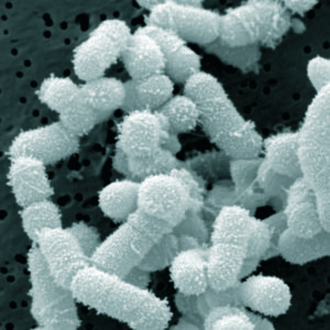
It's now 3 years being free of chronic sinusitis and off all antibiotics! Three amazing years since I started using easy do-it-yourself sinusitis treatments containing the probiotic (beneficial bacteria) Lactobacillus sakei. My sinuses feel great! And yes, it still feels miraculous.

After reading the original ground-breaking research on sinusitis done by Abreu et al (2012), it led to finding and trying L. sakei as a sinusitis treatment. Of course, there is an entire community of microbes that live in healthy sinuses (the sinus microbiome), but L. sakei seems to be a key one for sinus health.

I just updated the post The One Probiotic That Treats Sinusitis (originally posted January 2015) using my family's experiences (lots of self-experimentation!) and all the information that people have sent me. The post has a list of brands and products with L. sakei, as well as information about some other promising bacteria. Thank you so much! [For latest see: The Best Probiotic For Sinus Infections]

Thank you all who have written to me  - whether publicly or privately. Please keep writing and tell me what has worked or hasn't worked for you as a sinusitis treatment. If you find another bacteria or microbe or product that works for you - please let me know. It all adds to the sinusitis treatment knowledge base. I will keep posting updates. 

(NOTE: I wrote our background story - Sinusitis Treatment Story back in December 2013, and there is also a  Sinusitis Treatment Summary page with the various treatment methods. One can also click on SINUSITIS under CATEGORIES to see more posts, such as "Probiotics and Sinusitis" - a discussion by one of the original sinusitis researchers about what she thinks is going on in sinus microbiomes and what is needed.)  

This confirms what researchers such as Dr. Martin Blaser (in his book Missing Microbes) and others (such as Drs. Sonnenburg and Sonnenburg) have been saying about antibiotic use in infants and children: that there are negative effects to the gut microbiome from antibiotic use in early childhood, and the more frequent the use, the greater the negative effects. It is because the use of antibiotics  in early childhood "disrupts the microbiome".

Penicillins appear to be less disruptive, but macrolides (e.g., Clarithromycin, azithromycin) much more disruptive - the researchers found that the gut microbiota recovered within 6–12 months after a penicillin course, but did not fully recover from a macrolide course even after 2 years . Antibiotics can be life-saving, but they absolutely should not be used casually because there are hidden costs (such as microbiome changes). From Medical Xpress:

Antibiotic use in early life disrupt normal gut microbiota development

The use of antibiotics in early childhood interferes with normal development of the intestinal microbiota, shows research conducted at the University of Helsinki. Particularly the broad-spectrum macrolide antibiotics, commonly used to treat respiratory tract infections, have adverse effects. Macrolides appear also to contribute to the development of antibiotic-resistant strains of bacteria.  ...continue reading "Childhood Antibiotic Use Disrupts Gut Microbiome"

1

Once again, two opposing views about beards have been in the news - that they harbor all sorts of nasty disease-causing bacteria vs they are hygienic. An earlier May 5, 2015 post was about the question of whether bearded men have more bacteria on their faces than clean shaven ones. I cited a 2014 study found that they don't, and that we are all covered with bacteria, all sorts of bacteria, and this is normal.

Now another study has looked at the issue of hospital workers with and without beards and whether they carry infectious bacteria. Researchers swabbed the faces (center of the cheek and the skin of the upper lip under the nostrils) of both clean shaven individuals and individuals with facial hair (beards) that worked in two hospitals (they all had direct contact with patients) and looked at the bacteria present. They especially looked for the presence of the bacteria Staphylococcus aureus, which surprisingly was found more in the clean-shaven men.

Also to their surprise, it was more of the clean shaven men who carried the pathogenic bacteria Methicillin-resistant Staphylococcus aureus (also known as MRSA). For those bacterial groups most closely associated with hospital acquired infections, such as Klebsiella species, Pseudomonas species, Enterobacter species., and Acinetobacter species, prevalence was low in both groups, and less than 2% for each group.

For other, less harmful bacteria, researchers found that bearded employees harbored no more bacteria than their clean-shaven colleagues. In summary: The researchers say that "results suggest that male hospital workers with facial hair do not harbour more potentially concerning bacteria than clean-shaven workers, and that in some instances, clean-shaven individuals are significantly more likely to be colonized with potential nosocomial pathogens". (NOTE: nosocomial means a disease originating or acquired in a hospital.)

And why is that? According to the study, one explanation is "microtrauma to the skin," which occurs during shaving and results in abrasions, which could support bacterial colonisation and growth of bacteria on the clean-shaven men. However, some other researchers have a different hypothesis — that beards themselves actually fight infection.

This stems from an experiment carried out by Dr. Michael Mosley who recently swabbed the beards of a variety of men and sent the samples to Dr. Adam Roberts, a microbiologist at University College London. Roberts grew more than 100 different bacteria from the beard samples, but found that in a few of the petri dishes a microbe was killing the other bacteria -  a bacteria called Staphylococcus epidermidis, and which they believe has antibiotic properties.

From the Journal of Hospital Infection: Bacterial ecology of hospital workers’ facial hair: a cross-sectional study

Summary: It is unknown whether healthcare workers' facial hair harbours nosocomial pathogens. We compared facial bacterial colonization rates among 408 male healthcare workers with and without facial hair. Workers with facial hair were less likely to be colonized with Staphylococcus aureus (41.2% vs 52.6%, P = 0.02) and meticillin-resistant coagulase-negative staphylococci (2.0% vs 7.0%, P = 0.01). Colonization rates with Gram-negative organisms were low for all healthcare workers, and Gram-negative colonization rates did not differ by facial hair type. Overall, colonization is similar in male healthcare workers with and without facial hair; however, certain bacterial species were more prevalent in workers without facial hair.

[Excerpts from Discussion]:Several studies to date have demonstrated that physician white coats and neck ties can act as significant sources of nosocomial bacteria. Our study suggests that facial hair does not increase the overall risk of bacterial colonization compared to clean-shaven control subjects. Indeed, clean-shaven control subjects exhibited higher rates of colonization with certain bacterial species. This finding may be explained by microtrauma to the skin during shaving resulting in abrasions, which may support bacterial colonization and proliferation. This may be akin to the enhanced risk of surgical site infections in patients shaved with razors prior to surgery. Further, our results are consistent with prior evidence pertaining to bacterial colonization on the hands and nares of HCWs (Health care workers).