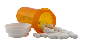
Skip to content

The finding that the oral bacteria Streptococcus mutans, which is found in 10% of the population, is linked with hemorrhagic strokes is big. S. mutans is found in tooth decay or cavities (dental caries). The researchers found a link with cnm-positive S. mutans with both intracerebral hemorrhage (ICH) and also with cerebral microbleeds.

Some risk factors for strokes have long since been known, such as high blood pressure and advanced age, but then there are those hemorrhagic strikes that don't seem to fit the norm, with no apparent risk factors. Well, apparently the presence of cnm-positive S. mutans is one. My understanding of what cnm-positive S. mutans means is S. mutans bacteria that carries the collagen-binding Cnm gene. This bacteria can be found in a person's saliva and in dental plaque, and swabs of both were taken for this study.

This study builds on other studies that find a link between the bacteria Streptococcus mutans and a number of systemic diseases, including bacteremia, infective endocarditis and hemorrhagic stroke. The researchers of this latest study suggest that infection with cnm-positive S. mutans causes constant inflammation (as shown by 2 inflammatory markers: CRP and fibrinogen), which then causes damage to blood vessels (endothelial damage) in the brain. Bottom line: take care of your teeth and gums.

From Science Daily: Oral bacteria linked to risk of stroke

In a study of patients entering the hospital for acute stroke, researchers have increased their understanding of an association between certain types of stroke and the presence of the oral bacteria (cnm-positive Streptococcus mutans).

In the single hospital study, researchers at the National Cerebral and Cardiovascular Center in Osaka, Japan, observed stroke patients to gain a better understanding of the relationship between hemorrhagic stroke and oral bacteria. Among the patients who experienced intracerebral hemorrhage (ICH), 26 percent were found to have a specific bacterium in their saliva, cnm-positive S. mutans. Among patients with other types of stroke, only 6 percent tested positive for the bacterium.

Strokes are characterized as either ischemic strokes, which involve a blockage of one or more blood vessels supplying the brain, or hemorrhagic strokes, in which blood vessels in the brain rupture, causing bleeding.

The researchers also evaluated MRIs of study subjects for the presence of cerebral microbleeds (CMB), small brain hemorrhages which may cause dementia and also often underlie ICH. They found that the number of CMBs was significantly higher in subjects with cnm-positive S. mutans than in those without. The authors hypothesize that the S. mutans bacteria may bind to blood vessels weakened by age and high blood pressure, causing arterial ruptures in the brain, leading to small or large hemorrhages.

"This study shows that oral health is important for brain health. People need to take care of their teeth because it is good for their brain and their heart as well as their teeth," Friedland said. "The study and related work in our labs have shown that oral bacteria are involved in several kinds of stroke, including brain hemorrhages and strokes that lead to dementia."

Multiple research studies have shown a close association between the presence of gum disease and heart disease, and a 2013 publication by Jan Potempa, Ph.D., D.Sc., of the UofL School of Dentistry, revealed how the bacterium responsible for gum disease worsens rheumatoid arthritisThe cnm-negative S. mutans bacteria is found in approximately 10 percent of the general population, Friedland says, and is known to cause dental cavities (tooth decay). Friedland also is researching the role of oral bacteria in other diseases affecting the brain.  http://www.nature.com/articles/srep20074

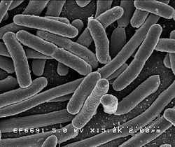
A recent study has examined the issue of whether the 10 to 1 ratio of bacteria to human cells, which is widely quoted, is actually correct. Weizmann Institute of Science researchers currently feel that based on scientific evidence (which of course will change over time) and making "educated estimates", the actual ratio is closer to 1:1 (but overall there still are more bacterial than human cells). They point out that the 10:1 ratio was originally a "back of the envelope" estimate dating back to 1972.

The researchers also point out that the ratio may vary over the course of each day - as a person defecates out huge amounts of bacteria with each bowel movement. However, this study - which is not the final word - is an educated guess about bacteria only. What about the viruses, the fungi, etc that also reside on and within us? We know much less about all the other microbes. I am disturbed that article after article, and headline after headline, equates microbes and bacteria. Microbes does not mean only bacteria.  From Science Daily:

Germs, humans and numbers: New estimate revises our microbiome numbers downwards

How many microbes inhabit our body on a regular basis? For the last few decades, the most commonly accepted estimate in the scientific world puts that number at around ten times as many bacterial as human cells. In research published in the journal Cell, a recalculation of that number by Weizmann Institute of Science researchers reveals that the average adult has just under 40 trillion bacterial cells and about 30 trillion human ones, making the ratio much closer to 1:1.

The rising importance of the microbiome in current scientific research led the Weizmann Institute's Prof. Ron Milo, Dr. Shai Fuchs and research student Ron Sender to revisit the common wisdom concerning the ratio of "personal" bacteria to human cells.

The original estimate that bacterial cells outnumber human cells in the body by ten to one was based on, among other things, the assumption that the average bacterium is about 1,000 times smaller than the average human cell. The problem with this estimate is that human cells vary widely in size, as do bacteria. For example, red blood cells are at least 100 times smaller than fat or muscle cells, and the microbes in the large intestine are about four times the size of the often-used "standard" bacterial cell volume. The Weizmann Institute scientists weighted their computations by the numbers of the different-sized human cells, as well as those of the various microbiome cells. 

Some excerpts from the original journal article from Cell: Are We Really Vastly Outnumbered? Revisiting the Ratio of Bacterial to Host Cells in Humans

The human microbiome has emerged as an area of utmost interest....One of the most fundamental and commonly cited figures in this growing field is the estimate that bacteria residing in the human body outnumber human cells by a factor of 10 or more (Figure 1A). This striking statement often serves as an entry point to the field. After all, if a human being is a cell population composed of at least 90% bacteria, it is only natural to expect a major role for them in human physiology.

Both the numerator (number of microbial cells) and the denominator (human cells) of this 10:1 ratio are based on crude assessments. Most sources cite the number of human cells as 1013 or 1014.....We performed a thorough review of the literature and found a long chain of citations originating from one “back of the envelope” estimate (Figure 1). This estimate, though illuminating, was never meant as the final word on the question.

Recently, the estimate of a 10:1 bacterial to human cell ratio (B/H) ratio has received criticism (Rosner, 2014). Therefore, an alternative value and an estimate of the uncertainty range are needed. Bacteria are found in many parts of the human body primarily on the external and internal surfaces, including the gastrointestinal tracts, skin, saliva, oral mucosa, and conjunctiva. The vast majority of commensal bacteria reside in the colon, with previous estimates of about 1014 bacteria (Savage, 1977), followed by the skin, which is estimated to harbor ∼1012 bacteriaBerg, 1996). Less than 1012 bacteria populate the rest of the body.....Almost all recent papers in the field of gut microbiota directly or indirectly rely on a single paper (Savage, 1977) discussing the overall number of bacteria in the gut. Interestingly, review of the original Savage 1977 paper demonstrates that it actually cites another paper for the estimate (Luckey, 1972)....The estimate, performed by Luckey in 1972, is an illuminating example of a back-of-the-envelope estimate, which was elegantly performed, yet was probably never meant to serve as the cornerstone reference number to be cited decades later.

Updating the ratio of bacteria to human cells from 10:1 or 100:1 to closer to 1:1 does not take away from the biological importance of the microbiota. ...Although we still appear to be outnumbered, we now know more reliably to what degree and can quantify our uncertainty about the ratios and absolute numbers. The B/H ratio is actually close enough to one, so that each defecation event, which excretes about 1/3 of the colonic bacterial content, may flip the ratio to favor human cells over bacteria. This anecdote serves to highlight that some variation in the ratio of bacterial to human cells occurs not only across individual humans but also over the course of the day.

I posted about this amazing research while it was still ongoing (Jan. 16, 2015), but now a study has been published. The small well-done pilot study looked at the microbiome (microbial communities) and microbial differences between different groups of infants during the first 30 days of life. They found significant differences in the bacteria of C-section infants (not exposed to their mother's vaginal fluid in the birth canal) compared to C-section infants who were swabbed with a gauze pad right after birth with their mother's vaginal fluids. They found that the microbiota (community of microbes) is partially restored in the swabbed C-section infants and more similar to that of vaginally delivered infants (who were exposed to the maternal bacteria naturally in the birth canal). They found that the procedure restored some bacteria, such as Lactobacillus and Bacteroides, which were nearly absent in the skin and anal samples of non-swabbed C-section babies.

In the C-section group, four mothers who were free of infections that might harm the babies, incubated a sterile gauze in their vaginas for one hour before the operation (C-section). Then, within two minutes of birth, the babies were swabbed with the gauze first over their mouths, then their faces, and then the rest of their bodies. These results are important because it is thought that microbiome differences (depending on method of birth) are long-lasting (with higher incidence of some health problems later in life with C-sections), and because the baby's early microbiome helps educate the baby's developing immune system.

Rob Knight (a leading microbiologist and one of the researchers) pointed out that the study "provides the proof-of-concept that microbiome modification early in life is possible." Now we need to see if these microbial differences persist over time and if it makes a health difference. From Science Daily:

Vaginal microbes can be partially restored to c-section babies

In a small pilot study, researchers at University of California, San Diego School of Medicine and Icahn School of Medicine at Mount Sinai determined that a simple swab to transfer vaginal microbes from a mother to her C-section-delivered newborn can alter the baby's microbial makeup (microbiome) in a way that more closely resembles the microbiome of a vaginally delivered baby. 

Babies delivered by C-section differ from babies delivered vaginally in the makeup of the microbes that live in and on their bodies. These early microbiomes help educate the baby's developing immune system. Previous research suggests a link between C-section delivery and increased subsequent risk of obesity, asthma, allergies, atopic disease and other immune deficiencies. Many of these diseases have also been linked to the microbiome, though the role a newborn's microbiome plays in current or long-term health is not yet well-understood....Other research suggests that microbiome differences between vaginal and C-section babies can persist for years."

In the study, the researchers collected samples from 18 infants and their mothers, including seven born vaginally and 11 delivered by scheduled C-section. Of the C-section-delivered babies, four were exposed to their mothers' vaginal fluids at birth as part of this study. To do this, sterile gauze was incubated in the mothers' vaginas for one hour before the C-section. Within two minutes of their birth, the babies delivered by C-section were swabbed with the gauze starting with the mouth, then the face and the rest of the body.

Six times over the first month after birth, the researchers collected a total of 1,519 anal, oral and skin samples from the mothers and infants. Knight's team then used a gene sequencing technique to map the types and relative quantities of bacterial species present at each body site.

Here's what they found: the microbiomes of the four C-section-delivered infants exposed to vaginal fluids more closely resembled those of vaginally delivered infants than unexposed C-section-delivered infants, though the difference was more distinct in their oral and skin samples than in their anal samples. This partial microbial restoration could be due to the fact that the infants received only one surface application of maternal vaginal fluids, Knight said.

Yet the oral and skin microbiome differences between C-section-delivered infants who received the microbial transfer and those who did not was still noticeable one month after birth. The results were not due to diet differences, as all of the infants received breast milk either exclusively or supplemented with formula during the first month of life. In addition, consistent with previous studies, the babies' microbiome profiles did not correlate with the amount of breast milk they received.

"The present work is a pilot study -- we need substantially more children and a longer follow-up period to connect the procedure to health effects," said Knight...."This study points the way to how we would do that, and provides the proof-of-concept that microbiome modification early in life is possible. In fact, we already have more than 10,000 additional samples collected as part of this study that still await analysis."

4

It's now 3 years being free of chronic sinusitis and off all antibiotics! Three amazing years since I started using easy do-it-yourself sinusitis treatments containing the probiotic (beneficial bacteria) Lactobacillus sakei. My sinuses feel great! And yes, it still feels miraculous.

After reading the original ground-breaking research on sinusitis done by Abreu et al (2012), it led to finding and trying L. sakei as a sinusitis treatment. Of course, there is an entire community of microbes that live in healthy sinuses (the sinus microbiome), but L. sakei seems to be a key one for sinus health.

I just updated the post The One Probiotic That Treats Sinusitis (originally posted January 2015) using my family's experiences (lots of self-experimentation!) and all the information that people have sent me. The post has a list of brands and products with L. sakei, as well as information about some other promising bacteria. Thank you so much! [For latest see: The Best Probiotic For Sinus Infections]

Thank you all who have written to me  - whether publicly or privately. Please keep writing and tell me what has worked or hasn't worked for you as a sinusitis treatment. If you find another bacteria or microbe or product that works for you - please let me know. It all adds to the sinusitis treatment knowledge base. I will keep posting updates. 

(NOTE: I wrote our background story - Sinusitis Treatment Story back in December 2013, and there is also a  Sinusitis Treatment Summary page with the various treatment methods. One can also click on SINUSITIS under CATEGORIES to see more posts, such as "Probiotics and Sinusitis" - a discussion by one of the original sinusitis researchers about what she thinks is going on in sinus microbiomes and what is needed.)  

This confirms what researchers such as Dr. Martin Blaser (in his book Missing Microbes) and others (such as Drs. Sonnenburg and Sonnenburg) have been saying about antibiotic use in infants and children: that there are negative effects to the gut microbiome from antibiotic use in early childhood, and the more frequent the use, the greater the negative effects. It is because the use of antibiotics  in early childhood "disrupts the microbiome".

Penicillins appear to be less disruptive, but macrolides (e.g., Clarithromycin, azithromycin) much more disruptive - the researchers found that the gut microbiota recovered within 6–12 months after a penicillin course, but did not fully recover from a macrolide course even after 2 years . Antibiotics can be life-saving, but they absolutely should not be used casually because there are hidden costs (such as microbiome changes). From Medical Xpress:

Antibiotic use in early life disrupt normal gut microbiota development

The use of antibiotics in early childhood interferes with normal development of the intestinal microbiota, shows research conducted at the University of Helsinki. Particularly the broad-spectrum macrolide antibiotics, commonly used to treat respiratory tract infections, have adverse effects. Macrolides appear also to contribute to the development of antibiotic-resistant strains of bacteria.  ...continue reading "Childhood Antibiotic Use Disrupts Gut Microbiome"

1

Once again, two opposing views about beards have been in the news - that they harbor all sorts of nasty disease-causing bacteria vs they are hygienic. An earlier May 5, 2015 post was about the question of whether bearded men have more bacteria on their faces than clean shaven ones. I cited a 2014 study found that they don't, and that we are all covered with bacteria, all sorts of bacteria, and this is normal.

Now another study has looked at the issue of hospital workers with and without beards and whether they carry infectious bacteria. Researchers swabbed the faces (center of the cheek and the skin of the upper lip under the nostrils) of both clean shaven individuals and individuals with facial hair (beards) that worked in two hospitals (they all had direct contact with patients) and looked at the bacteria present. They especially looked for the presence of the bacteria Staphylococcus aureus, which surprisingly was found more in the clean-shaven men.

Also to their surprise, it was more of the clean shaven men who carried the pathogenic bacteria Methicillin-resistant Staphylococcus aureus (also known as MRSA). For those bacterial groups most closely associated with hospital acquired infections, such as Klebsiella species, Pseudomonas species, Enterobacter species., and Acinetobacter species, prevalence was low in both groups, and less than 2% for each group.

For other, less harmful bacteria, researchers found that bearded employees harbored no more bacteria than their clean-shaven colleagues. In summary: The researchers say that "results suggest that male hospital workers with facial hair do not harbour more potentially concerning bacteria than clean-shaven workers, and that in some instances, clean-shaven individuals are significantly more likely to be colonized with potential nosocomial pathogens". (NOTE: nosocomial means a disease originating or acquired in a hospital.)

And why is that? According to the study, one explanation is "microtrauma to the skin," which occurs during shaving and results in abrasions, which could support bacterial colonisation and growth of bacteria on the clean-shaven men. However, some other researchers have a different hypothesis — that beards themselves actually fight infection.

This stems from an experiment carried out by Dr. Michael Mosley who recently swabbed the beards of a variety of men and sent the samples to Dr. Adam Roberts, a microbiologist at University College London. Roberts grew more than 100 different bacteria from the beard samples, but found that in a few of the petri dishes a microbe was killing the other bacteria -  a bacteria called Staphylococcus epidermidis, and which they believe has antibiotic properties.

From the Journal of Hospital Infection: Bacterial ecology of hospital workers’ facial hair: a cross-sectional study

Summary: It is unknown whether healthcare workers' facial hair harbours nosocomial pathogens. We compared facial bacterial colonization rates among 408 male healthcare workers with and without facial hair. Workers with facial hair were less likely to be colonized with Staphylococcus aureus (41.2% vs 52.6%, P = 0.02) and meticillin-resistant coagulase-negative staphylococci (2.0% vs 7.0%, P = 0.01). Colonization rates with Gram-negative organisms were low for all healthcare workers, and Gram-negative colonization rates did not differ by facial hair type. Overall, colonization is similar in male healthcare workers with and without facial hair; however, certain bacterial species were more prevalent in workers without facial hair.

[Excerpts from Discussion]:Several studies to date have demonstrated that physician white coats and neck ties can act as significant sources of nosocomial bacteria. Our study suggests that facial hair does not increase the overall risk of bacterial colonization compared to clean-shaven control subjects. Indeed, clean-shaven control subjects exhibited higher rates of colonization with certain bacterial species. This finding may be explained by microtrauma to the skin during shaving resulting in abrasions, which may support bacterial colonization and proliferation. This may be akin to the enhanced risk of surgical site infections in patients shaved with razors prior to surgery. Further, our results are consistent with prior evidence pertaining to bacterial colonization on the hands and nares of HCWs (Health care workers).

A recent study using mice, and following them for 4 generations, has implications for Americans who typically eat a low-fiber diet (average of 15 grams daily). Note that current dietary guidelines recommend that women should eat around 25 grams and men 38 grams daily of fiber. The researchers found that low-fiber diets not only deplete the complex microbial ecosystems residing in the gut, but can cause an irreversible loss of diversity within those ecosystems in as few as three or four generations.

This is because fiber feeds the millions of microbes in the gut - and so a fiber-rich diet can nourish a wide variety of gut microbes, but a low-fiber diet can only sustain a narrower community. As the generations went by, the rodents’ guts became progressively less diverse, as more and more species were extinguished. If the fourth-generation mice switched to high-fiber meals, some of the missing microbes rebounded, but most did not. It took a fecal transplant (mice style) to get back the missing microbes. From Science Daily:

Low-fiber diet may cause irreversible depletion of gut bacteria over generations

A study by Stanford University School of Medicine investigators raises concerns that the lower-fiber diets typical in industrialized societies may produce internal deficiencies that get passed along to future generations. The study, conducted in mice, indicates that low-fiber diets not only deplete the complex microbial ecosystems residing in every mammalian gut, but can cause an irreversible loss of diversity within those ecosystems in as few as three or four generations.

Once an entire population has experienced the extinction of key bacterial species, simply "eating right" may no longer be enough to restore these lost species to the guts of individuals in that population, the study suggests. Those of us who live in advanced industrial societies may already be heading down that path.

Bad news about ticks: the blacklegged ticks (Ixodes scapularis and the western Ixodes pacificus) that spread Lyme disease, are now reported in almost half of the counties in the U. S. Researchers found blacklegged ticks in 1,420 out of 3,110 counties in the continental U.S., or about 46% of counties, and found western blacklegged ticks in 111 counties, or about 4%. Combined, this is a 45% increase from 1998 when ticks were reported in 1,058 counties.Of course the tick-dense northeast is where Lyme disease is most common. Although the blacklegged tick is found from Florida to Minnesota, 95% of confirmed Lyme disease cases come from just 14 states in the northeast and upper Midwest. 

One interesting study that looked at reasons for these differences was published in PLOS ONE last year by parasitologist Isis Arsnoe and colleagues . They found that populations of blacklegged ticks behave differently in the north and the south United States. Nymphs of the blacklegged tick in the north are bolder and more active in seeking out hosts, a behavior known as questing. Arsnoe found that that tick nymphs originating from Wisconsin and Rhode Island were 20 times more likely to emerge from leaf litter, putting them in the path of passing humans, than nymphs from North or South Carolina. "Questing behavior is a key factor affecting the risk of tick bites." From Science Daily:

Ticks that transmit Lyme disease reported in nearly half of all US counties

Lyme disease is transmitted by the blacklegged tick (Ixodes scapularis) and the western blacklegged tick (Ixodes pacificus), and the range of these ticks is spreading, according to research published in the Journal of Medical Entomology.

Some symptoms of Lyme disease include fever, headache, and fatigue, all of which can be mistaken for the common flu, so medical personnel need to know where these ticks are found in order to make a correct diagnosis. Unfortunately, the range of blacklegged ticks had not been re-evaluated in nearly two decades, until now.

The team used surveillance methods similar to those used in 1998 so that they would be able to accurately judge the degree to which the distribution of these ticks had changed. Using the gathered data, they figured out which counties had established populations, which ones had one or more reports of a blacklegged ticks, and which ones had none.

They found that the blacklegged tick has been reported in more than 45% of  U.S. counties, compared to 30% of counties in 1998. Even more alarming, the blacklegged tick is now considered established in twice the number of counties as in 1998. Most of the geographic expansion of the blacklegged tick appears to be in the northern U.S., while populations in southern states have remained relatively stable. The range of the western blacklegged tick only increased from 3.4% to 3.6% of counties. (The study in J. of Medical Entomology)

The following article is interesting because it describes how microbes are high up in the sky riding air currents and winds to circle the earth, and eventually drop down somewhere. This is one way diseases can be spread from one part of the world to another. And the study looking at how antibiotic resistant bacteria are spread in the air from cattle feedlots has implications for how antibiotic resistance is spread. From Smithsonian:

Living Bacteria Are Riding Earth's Air Currents

Considering the prevailing winds, David J. Smith figured the air samples collected atop a dormant volcano in Oregon would be full of DNA signatures from dead microorganisms from Asia and the Pacific Ocean. He didn’t expect anything could survive the journey through the harsh upper atmosphere to the research station at the Mount Bachelor Observatory, at an elevation of 9,000 feet.

But when his team got to the lab with the samples, taken from two large dust plumes in the spring of 2011, they discovered a thriving bunch of hitchhikers. More than 27 percent of the bacterial samples and more than 47 percent of the fungal samples were still alive. Ultimately, the team detected about 2,100 species of microbes, including a type of Archea that had only previously been isolated off the coast of Japan. “In my mind, that was the smoking gun,“ Smith says. Asia, as he likes to say, had sneezed on North America.

 Microbes have been found in the skies since Darwin collected windswept dust aboard the H.M.S. Beagle 1,000 miles west of Africa in the 1830s. But technologies for DNA analysis, high-altitude collection and atmospheric modeling are giving scientists a new look at crowded life high above Earth. For instance, recent research suggests that microbes are hidden players in the atmosphere, making clouds, causing rain, spreading diseases between continents and maybe even changing climates.

"I regard the atmosphere as a highway, in the most literal sense of the term," Smith says. "It enables the exchange of microorganisms between ecosystems thousands of miles apart, and to me that’s a more profound ecological consequence we still have not fully wrapped our heads around."

Airborne microbes potentially have huge impacts on our planet. Some scientists attribute a 2001 foot-and-mouth outbreak in Britain to a giant storm in north Africa that carried dust and possibly spores of the animal disease thousands of miles north only a week before the first reported cases. Bluetongue virus, which infects domestic and wild animals, was once present only in Africa. But it's found now in Great Britain, likely the result of the prevailing winds.

In west Texas, researchers from Texas Tech University collected air samples upwind and downwind of ten cattle feedlots. Antibiotic resistant microbes were 4,000 percent more prevalent in the downwind samples. .... What's clear is there are far more viable microbes in far more inhospitable places than scientists expected.

New research that found that microbial communities vary between the sinuses in a person with chronic sinusitis. This is a result that many sinusitis sufferers already suspect based on their sinusitis symptoms. The researchers also found that bacterial communities in the sinuses vary between people with chronic sinusitis. It is frustrating though for me to read study after study where the researchers focus on describing the types of bacteria found in chronic sinusitis sufferers (and then just saying that the sinus microbiomes or community of microbes vary from person to person) rather than studies comparing the sinus microbiomes (bacteria and other microbes, such as fungi) between healthy individuals and sinusitis sufferers.

Since research finds that sinusitis sufferers have altered sinus microbiomes, then what would be really helpful now is finding more beneficial or keystone species (besides Lactobacillus sakei) that are needed for healthy sinus microbiomes. This would be an important step towards then adding (perhaps using a nasal spray) these missing microbes to the sinus microbiome. From Frontiers in Microbiology:

Bacterial communities vary between sinuses in chronic rhinosinusitis patients

ABSTRACT: Chronic rhinosinusitis (CRS) is a common and potentially debilitating disease characterized by inflammation of the sinus mucosa for longer than 12 weeks. Bacterial colonization of the sinuses and its role in the pathogenesis of this disease is an ongoing area of research. Recent advances in culture-independent molecular techniques for bacterial identification have the potential to provide a more accurate and complete assessment of the sinus microbiome, however there is little concordance in results between studies, possibly due to differences in the sampling location and techniques. This study aimed to determine whether the microbial communities from one sinus could be considered representative of all sinuses, and examine differences between two commonly used methods for sample collection, swabs and tissue biopsies. High-throughput DNA sequencing of the bacterial 16S rRNA gene was applied to both swab and tissue samples from multiple sinuses of 19 patients undergoing surgery for treatment of CRS. Results from swabs and tissue biopsies showed a high degree of similarity, indicating that swabbing is sufficient to recover the microbial community from the sinuses. Microbial communities from different sinuses within individual patients differed to varying degrees, demonstrating that it is possible for distinct microbiomes to exist simultaneously in different sinuses of the same patient. The sequencing results correlated well with culture-based pathogen identification conducted in parallel, although the culturing missed many species detected by sequencing. This finding has implications for future research into the sinus microbiome, which should take this heterogeneity into account by sampling patients from more than one sinus. It may also be of clinical importance, as determination of antibiotic sensitivities using culture of a swab from a single sinus could miss relevant pathogens that are localized to another sinus.

CRS can be a debilitating condition that is recalcitrant to treatment. Bacterial colonization of the sinuses is likely to play an important role in the pathogenesis and perpetuation of the disease; however different studies have yielded contrasting results with respect to which bacterial taxa are characteristic of the disease (ref). We observed bacterial communities dominated by different taxa in CRS patients; for example some have sinuses colonized primarily with Haemophilus, while others are dominated by Corynebacterium and Staphylococcus, or Pseudomonas. Some patients’ sinuses contain anaerobic bacteria such as Anaerococcus, Finegoldia, and Peptoniphilus, while these were absent from others. Indeed, our results have shown, for the first time, that it is possible for a patient to simultaneously have different bacterial communities in different sinuses, pointing to distinct, localized microbiomes within the same patient. Understanding this variation in the sinus microbiome could prove critical to the appropriate selection of treatments for CRS in the future.

The weighted unifrac distances between samples within patients (Figure 1) demonstrate that at least some CRS patients have substantial variation of bacterial communities between sinuses, although it is significantly smaller than the variation observed between different individuals. While this variation was related to abundance rather than the presence or absence of dominant community members, some of these variations were large: for example Corynebacterium sequences dominating the right sinuses of patient 003 (60.7 and 41.7% of all sequences), while the left sinuses had much smaller abundances (9.8 and 6.2%) and were dominated by the anaerobic bacteria Anaerococcus, Finegoldia  and Peptinophillus.