 A study by researchers showing troubling effects from certain pesticides (especially a class of fungicides) raises all sorts of questions: What is the long-term effect of chronic low doses of these fungicides in the foods we eat? How much of these chemicals are we getting exposed to? The Univ. of North Carolina researchers studied the effect of 294 chemicals (all common food-use pesticides or other environmental chemicals) on "mouse cortical neurons" (mouse brain cells). They found that one group of chemicals, which they referred to as "cluster 2", "mimics brain disorders" such as autism, advanced age, Alzheimer's, Parkinson's disease, and other neurodegenerative disorders. The chemicals (all pesticides, and mainly fungicides) causing these effects are: fenamidone, pyraclostrobin, famoxadone, trifloxystrobin, fenpyroximate, azoxystrobin, fluoxastrobin pyridaben and rotenone. Even though this study was done on mouse cortical neurons (in vitro), it is meaningful because of the similarities with human brain cells.
Very little is known about human exposure to these chemicals (how much is our exposure?) and their effects on humans, but the data suggest effects similar to that in neurological disorders. The researchers point out that many of the chemical residues in this cluster were found on conventionally raised foods, especially leafy green vegetables, and were detected at relatively high levels, especially pyraclostrobin. Most of these fungicides only came into use after 2000 and usage of these fungicides has been increasing in the U.S, with the exception of pyridaben (decreasing use) and rotenone (very low use). "These data suggest significant human exposure potential to many of the chemicals in cluster 2".
They point out that these fungicide residues have not been detected on organically produced foods (EPA and USDA data), which suggests a way to minimize exposure. None of these chemicals can be used by organic farmers in the U.S. Possible exposure is also from gardens and lawns (if used), contaminated water, and for farm workers in conventional agriculture. From Science Daily:
Could new class of fungicides play a role in autism, neurodegenerative diseases?
Scientists at the UNC School of Medicine have found a class of commonly used fungicides that produce gene expression changes similar to those in people with autism and neurodegenerative conditions, including Alzheimer's disease and Huntington's disease.
Mark Zylka, PhD, senior author of the study and associate professor of cell biology and physiology at UNC, and his team exposed mouse neurons to approximately 300 different chemicals.... "Based on RNA sequencing, we describe six groups of chemicals," Zylka said. "We found that chemicals within each group altered expression in a common manner. One of these groups of chemicals altered the levels of many of the same genes that are altered in the brains of people with autism or Alzheimer's disease." Chemicals in this group included the pesticides rotenone, pyridaben, and fenpyroximate, and a new class of fungicides that includes pyraclostrobin, trifloxystrobin, fenamidone, and famoxadone. Azoxystrobin, fluoxastrobin, and kresoxim-methyl are also in this fungicide class.
"We cannot say that these chemicals cause these conditions in people," Zylka cautioned. "Many additional studies will be needed to determine if any of these chemicals represent real risks to the human brain." Zylka, a member of the UNC Neuroscience Center, and his group found that these chemicals reduced the expression of genes involved in synaptic transmission -- the connections important for communication between neurons. If these genes are not expressed properly, then our brains cannot function normally. Also, these chemicals caused an elevated expression of genes associated with inflammation in the nervous system. This so-called neuroinflammation is commonly seen in autism and neurodegenerative conditions.
The researchers also found that these chemicals stimulated the production of free radicals -- particles that can damage the basic building blocks of cells and that have been implicated in a number of brain diseases. The chemicals also disrupted neuron microtubules. "Disrupting microtubules affects the function of synapses in mature neurons and can impair the movement of cells as the brain develops," Zylka said. "We know that deficits in neuron migration can lead to neurodevelopmental abnormalities. We have not yet evaluated whether these chemicals impair brain development in animal models or people."
Jeannie T. Lee, MD, PhD, professor of genetics at Harvard Medical School and Massachusetts General Hospital, who was not involved in this research, said, "This is a very important study that should serve as a wake-up call to regulatory agencies and the general medical community. The work is timely and has wide-ranging implications not only for diseases like autism, Parkinson's, and cancer, but also for the health of future generations. I suspect that a number of these chemicals will turn out to have effects on transgenerational inheritance."
Zylka's group also analyzed information from the U.S. Geological Survey, which monitors countywide pesticide usage, as well as the Food and Drug Administration and the U.S. Department of Agriculture, which test foodstuffs yearly for pesticide residues. Of the chemicals Zylka's team studied, only the usage of pyridaben has decreased since 2000. Rotenone use has remained the same since 2000. However, the use of all the fungicides in this group has increased dramatically over the past decade.
Indeed, a study from the Environmental Protection Agency found that pyraclostrobin is found on foods at levels that could potentially affect human biology, and another study linked pyraclostrobin usage to honeybee colony collapse disorder. The pesticide rotenone was previously implicated in Parkinson's disease through replicated animal experiments and through human epidemiological studies.....Previous work has also shown that a single dose of the fungicide trifloxystrobin reduced motor activity for several hours in female rats and for days in male rats. Disrupted motor function is a common symptom of Parkinson's disease and other neurological disorders. The related fungicide picoxystrobin impaired motor activity in rats at the lowest dose tested.
Zylka added, "The real tough question is: if you eat fruits, vegetables or cereals that contain these chemicals, do they get into your blood stream and at what concentration? That information doesn't exist." Also, given their presence on a variety of foodstuffs, might long term exposure to these chemicals -- even at low doses -- have a cumulative effect on the brain?
Zylka noted that conventionally grown leafy green vegetables such as lettuce, spinach, and kale have the highest levels of these fungicides. But due to each chemical's effectiveness at reducing fungal blights and rust, crop yields have increased and farmers are expanding their use of these chemicals to include many additional types of food crops.
Zylka's team hopes their research will encourage other scientists and regulatory agencies to take a closer look at these fungicides and follow up with epidemiological studies. "Virtually nothing is known about how these chemicals impact the developing or adult brain," Zylka said. "Yet these chemicals are being used at increasing levels on many of the foods we eat."
 Applying fungicide to apple orchard. Credit: Univ. of Kentucky Agriculture Extension
 Another study that found benefits to dog ownership. The study authors concluded that: "Our study provides evidence that dog owners are at a lower risk for ischemic stroke, hemorrhagic stroke and heart failure." This could be to daily exercise, or that dog ownership results in less stress or better psychosocial health, or even some other reason (perhaps dog owners are healthier to start with). Note: myocardial infarction (MI) is commonly known as a heart attack. From Medscape:
 Toxoplasma gondii tissue cyst in a mouse brain
 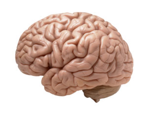 Another study showing that higher physical activity (from a variety of activities) is "related to larger gray matter volume in the elderly, regardless of cognitive status", specifically in gray matter areas of the brain responsible for memory, learning, and cognition. In other words, higher levels of physical activity reduce brain atrophy that occurs with aging and improves cognitive function in elderly individuals. There is also discussion of higher activity levels improving cerebral (brain) blood flow. Bottom line: get off your butt and move more for better brain health. From Medical Xpress:
 A new study has confirmed an association between proton pump inhibitors (PPIs) — drugs that treat heartburn, peptic ulcers, and other acid-related disorders of the upper gastrointestinal tract — and increased risk for dementia in older patients. An earlier study by the same researchers found the same link between PPI use and dementia risk. The drugs work by lowering the amount of acid produced by the stomach. PPIs are among the most frequently prescribed drugs, and include omeprazole (Losec), esomeprazole (Nexium), lansoprazole (Prevacid), and the over-the-counter medication Olex.
 The finding that the oral bacteria Streptococcus mutans, which is found in 10% of the population, is linked with hemorrhagic strokes is big. S. mutans is found in tooth decay or cavities (dental caries). The researchers found a link with cnm-positive S. mutans with both intracerebral hemorrhage (ICH) and also with cerebral microbleeds.
 More evidence that traditional toys and books are superior to electronic toys in both verbal parent-child interactions and non-verbal interactions for young children. Parent-child verbal interactions are so important because they teach young children language, lay the groundwork for literacy skills, teach role-playing, teach emotional and social skills such as turn-taking and accepting others' leads. In other words, put down the electronic gadgets and go spend time talking and interacting with your young child with old style traditional toys and books. From Science Daily: