Skip to content

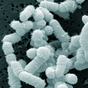
An interesting study that compared bacterial communities between healthy children and those that have a history of acute sinusitis (but not chronic sinusitis). The study specifically looked at the nasopharyngeal (NP)  microbiome (community of microbes) over the course of one year in the 2 groups of children, who were between the ages of 4 and 7. Nasopharyngeal pertains to the nose or nasal cavity and pharynx. They used modern methods of genetic analysis to test for bacterial species - and found a total of 951 species among the 47 children, of which 308 species had some "depletion" among those children with a history of sinusitis, and one species was increased in "abundance".

NP samples from children with a prior history of acute sinusitis were characterized by significant depletion of bacterial species, including those in the Akkermansia, Faecalibacterium prausnitzii, Clostridium, Lactobacillus, Prevotella, and Streptococcus species. But there was a siignificant increase "in relative abundance" in the bacterial species Moraxella nonliquefaciens. Once again, a study shows bacterial communities to be "out of whack" in those who've had sinusitis - this time in children. And the diminished diversity was linked to more frequent upper respiratory illnesses. The researchers mention the "possibility that the manipulation of the airway microbiota" could help prevent childhood respiratory diseases. Research by C.A. Santee et al from the Microbiome journal at BioMed Central:

Nasopharyngeal microbiota composition of children is related to the frequency of upper respiratory infection and acute sinusitis

Upper respiratory infections (URI) and their complications are a major healthcare burden for pediatric populations. Although the microbiology of the nasopharynx is an important determinant of the complications of URI, little is known of the nasopharyngeal (NP) microbiota of children, the factors that affect its composition, and its precise relationship with URI.

Healthy children (n = 47) aged 49–84 months from a prospective cohort study based in Wisconsin, USA, were examined. Demographic and clinical data and NP swab samples were obtained from participants upon entry to the study. All NP samples were profiled for bacterial microbiota using a phylogenetic microarray, and these data were related to demographic characteristics and upper respiratory health outcomes. The composition of the NP bacterial community of children was significantly related prior to the history of acute sinusitisHistory of acute sinusitis was associated with significant depletion in relative abundance of taxa including Faecalibacterium prausnitzii and Akkermansia spp. and enrichment of Moraxella nonliquefaciens. Enrichment of M. nonliquefaciens was also a characteristic of baseline NP samples of children who subsequently developed acute sinusitis over the 1-year study period. Time to develop URI was significantly positively correlated with NP diversity, and children who experienced more frequent URIs exhibited significantly diminished NP microbiota diversity (P ≤ 0.05). 

These preliminary data suggest that previous history of acute sinusitis influences the composition of the NP microbiota, characterized by a depletion in relative abundance of specific taxa. Diminished diversity was associated with more frequent URIs

....These observations indicate that the composition of the pediatric upper airway represents a critical factor that may either potentiate or protect against infection by respiratory pathogens. They also indicate that the interplay between the bacterial microbiota and respiratory pathogens associated with upper airway infection is important to consider.Both bacteria and viruses can influence each other’s pathogenicity [8] and a number of interactions between specific viruses and bacterial species have been reported in the airways [910]. For example, human rhinovirus infection was found to significantly increase the binding of Staphylococcus aureus, S. pneumoniae, or H. influenzae to primary human nasal epithelial cells [11]....

A total of 951 taxa were identified in baseline NP microbiota of participants (n = 47) in our cohort. These bacterial communities were variably composed of members of the Rickenellaceae, Lachnospiraceae, Verrucomicrobiaceae, Pseudomonadaceae, and Moraxellaceae as well as multiple unclassified members of the phylum Proteobacteria. .... Our study used independent NP samples collected from individual participants over a 12-month study period that spanned all four seasons. Season of sample collection also demonstrated a relationship with bacterial beta-diversity.

Compared with children who had no history of acute sinusitis (n = 33), those with a past history of acute sinusitis (n = 14) did not exhibit differences in α-diversity indices, suggesting that differences in microbiota characterizing these groups may be due to the enrichment or depletion of a subset of taxa within these bacterial communities. A total of 309 taxa (representing 101 genera) exhibited significant differences in relative abundance between children with and without a history of acute sinusitis. NP samples from children with a prior history of acute sinusitis were characterized by significant depletion of 308 of the 309 taxa, including those represented by Akkermansia, Faecalibacterium prausnitzii, Clostridium, Lactobacillus, Prevotella, and Streptococcus species. The only taxon that exhibited a significant increase in relative abundance in these subjects was represented by Moraxella nonliquefaciens. 

Children who experienced at least one URI (n = 17) within 60 days of collection of the baseline sample had significantly lower phylogenetic diversity compared to those who had no URIs within that time frame (n = 23). Time to development of URI, defined as the number of days between the collection of the baseline sample and the first incidence of URI (a value of 365 days was assigned to those children who did not experience a URI during the year of monitoring), was also significantly correlated with phylogenetic diversity .... Hence, these data indicate that diminished diversity of the NP microbiota is a precursor to URI in these children.  

In addition to Moraxella, a Corynebacterium was enriched in relative abundance in the NP microbiota of children who experienced acute sinusitis subsequent to baseline sample collection during the study period. ... However, Abreu et al. previously found Corynebacterium tuberculostearicum to be significantly enriched in the maxillary sinuses of adults with chronic rhinosinusitis compared to healthy control subjects [17]. The authors subsequently confirmed the ability of C. tuberculostearicum to induce acute sinusitis in the context of an antimicrobial-depleted murine model of sinus infection. Moreover co-installation of Lactobacillus sakei (one of a number of taxa acutely depleted in relative abundance among chronic rhinosinusitis patients) protected animals against C. tuberculostearicum infection [17]. Our pediatric data exhibits similarity with these murine studies, in that six members of the Lactobacillus genus were among those taxa most significantly depleted in relative abundance in the NP bacterial communities of children who developed sinusitis during our study. Five of these same taxa were also depleted in relative abundance in the NP microbial communities of children with a prior history of sinusitis. 

In addition to Lactobacillus, many other bacterial taxa including Akkermansia, Faecalibacterium prausnitzii, Clostridium, Prevotella, and Streptococcus species were depleted in relative abundance among children with a prior history of acute sinusitis. Though traditionally associated with gut microbiota, anaerobic bacterial species can exist in biofilms in the upper respiratory tract [18] and Akkermansia  and Faecalibacterium have previously been detected in the nasopharynx of children [1920]. While its role in the airway is unknown, gastrointestinal Akkermansia muciniphilia metabolizes mucin and has been shown to activate immune homeostasis, increasing host expression of antimicrobial peptides such as RegIIIγand improving barrier function via an increase in 2-oleoylgylcercerol [212223]. However, whether such mechanisms play a role at the airway mucosal surface remains to be determined. 

Mechanisms by which Lactobacillus and other bacterial species depleted in the NP microbiota of sinusitis patients may prevent the development of disease include competitive exclusion of pathogenic species. A previous murine study indicated that intra-nasal inoculation of mice with L. fermentum decreased S. pneumoniae burden throughout the respiratory tract and increased the number of activated macrophages in the lung and lymphocytes in the tracheal lamina propria [24]. Hence, it is plausible that the absence of NP genera with known competitive exclusion and immunomodulatory capabilities leads to pathogen expansion and associated clinical manifestations of upper airway infection. 

....We do show that a history of sinusitis, its pathophysiology or treatment, may shape the NP microbiota—which may inform future studies and their design. Additionally, though we recognize that the composition of the microbiota in the upper airways is likely highly influenced by antibiotic administration .... The pervasive effects of antimicrobials on the human microbiota are well-described [2627], and it is likely that lifetime antibiotic use plays an important role in shaping the baseline NP microbial community

The composition of the NP microbiota in healthy children between 49 and 84 months of age is associated with past and subsequent history of acute sinusitis and frequency of URI. Widespread bacterial taxon depletion and enrichment of M. liquefaciens and C. tuberculostearicum are associated with upper airway infection and the development of acute sinusitis. Collectively, these findings provide evidence of close connections between microbial colonization of the airways and susceptibility to upper respiratory illnesses in early childhood and raise the possibility that the manipulation of the airway microbiota could be applied to the prevention of childhood respiratory illnesses. 

Another excellent reason to lose weight if you are overweight or obese: losing weight (through diet or through combined diet and exercise) significantly lowers levels of proteins in the blood that help cancerous tumors grow. In other words, reducing weight could turn out to be a cancer prevention method in overweight and obese persons. Exercise alone did not lower the levels of these cancer-associated proteins.

The study enrolled 439 overweight or obese women (aged 50 to 75 years old) from the Seattle area who were randomly placed into one of four groups for 12 months: exercise only, diet only, exercise plus diet, or no change to health habits. Researchers measured three proteins in blood samples - VEGF, PAI-1 and PEDF – that flow through the body and help in the formation of new blood vessels, a process called angiogenesis. Angiogenesis can occur during such processes as wound healing, but it also occurs during the growth of tumors. Since the three measured proteins are involved in nurturing the growth and survival of tumor cells, this is a great reason to lose weight - to lower their levels in the blood. From Science Daily:

Losing weight lowered levels of proteins associated with tumor growth

Overweight or obese women who lost weight through diet or a combination of diet and exercise also significantly lowered levels of proteins in the blood that help certain tumors grow, according to a Fred Hutchinson Cancer Research Center study published July 14 in Cancer Research, a journal of the American Association for Cancer Research.

The study: Measured three proteins that are known to enhance tumor-related angiogenesis -- the formation of blood vessels that feed tumors and enable them to grow. Was intended to see how cancer-promoting proteins changed when overweight, sedentary, postmenopausal women lost weight through diet or diet and exercise over the course of a year. Enrolled 439 healthy women (they did not have cancer), placing each participant in one of four study arms: 1) Calorie- and fat-restricted diet. 2) Aerobic exercise five days a week. 3) Combined diet and exercise. 4) Control (no intervention).

Found that women in the diet arm and the diet and exercise arm lost more weight and had significantly lower levels of angiogenesis-related proteins, compared with women in the exercise-only arm and the control arm.

This study shows that weight loss may be a safe and effective way to improve the "angiogenic profile" of healthy individuals, meaning they would have lower blood levels of cancer-promoting proteins. Although the researchers cannot say for certain that this would impact the growth of tumors, they believe there could be an association between reduced protein levels and a less favorable environment for tumor growth.

Yup, according to a new mega-study, being overweight or obese is linked to higher risk of dying prematurely than being normal weight.  And the more you weigh, the greater the risk. This mega-study that looked at data from many studies and countries, also found that being underweight is linked to a higher risk of premature death. What's the best weight to be? A BMI of 22.5-<25 kg/m2 is considered a healthy weight range, and had the lowest mortality risk in the study. Being overweight was linked to higher rates of death from "all causes", and also from 4 major causes: coronary heart disease, stroke, respiratory disease, and cancer.

However, note that while other studies also agree that being underweight or obese increases the rate of dying prematurely, there is still some debate over whether being just overweight with BMI 25–<30 kg/m2 , really has a higher risk of dying prematurely. This was pointed out in the accompanying editorial in the journal Lancet (but not mentioned below). From Science Daily:

As overweight and obesity increase, so does risk of dying prematurely: Major study

Being overweight or obese is associated with a higher risk of dying prematurely than being normal weight -- and the risk increases with additional pounds, according to a large international collaborative study led by researchers at the Harvard T.H. Chan School of Public Health and the University of Cambridge, UK. The findings contradict recent reports that suggest a survival advantage to being overweight -- the so-called "obesity paradox."

The deleterious effects of excess body weight on chronic disease have been well documented. Recent studies suggesting otherwise have resulted in confusion among the public about what is a healthy weight. According to the authors of the new study, those prior studies had serious methodological limitations. One common problem is called reverse causation, in which a low body weight is the result of underlying or preclinical illness rather than the cause. Another problem is confounding by smoking because smokers tend to weigh less than nonsmokers but have much higher mortality rates.....Hu stressed that doctors should continue to counsel patients regarding the deleterious effects of excess body weight, which include a higher risk of diabetes, cardiovascular disease, and cancer.

For the new study, consortium researchers looked at data from more than 10.6 million participants from 239 large studies, conducted between 1970 and 2015, in 32 countries. A combined 1.6 million deaths were recorded across these studies, in which participants were followed for an average of 14 years. For the primary analyses, to address potential biases caused by smoking and preexisting diseases, the researchers excluded participants who were current or former smokers, those who had chronic diseases at the beginning of the study, and any who died in the first five years of follow-up, so that the group they analyzed included 4 million adults. They looked at participants' body mass index (BMI) -- an indicator of body fat calculated by dividing a person's weight in kilograms by their height in meters squared (kg/m2).

The results showed that participants with BMI of 22.5-<25 kg/m2 (considered a healthy weight range) had the lowest mortality risk during the time they were followed. The risk of mortality increased significantly throughout the overweight range: a BMI of 25-<27.5 kg/m2 was associated with a 7% higher risk of mortality; a BMI of 27.5-<30 kg/m2 was associated with a 20% higher risk; a BMI of 30.0-<35.0 kg/m2 was associated with a 45% higher risk; a BMI of 35.0-<40.0 kg/m2 was associated with a 94% higher risk; and a BMI of 40.0-<60.0 kg/m2 was associated with a nearly three-fold risk. Every 5 units higher BMI above 25 kg/m2 was associated with about 31% higher risk of premature death. Participants who were underweight also had a higher mortality risk.

Looking at specific causes of death, the study found that, for each 5-unit increase in BMI above 25 kg/m2, the corresponding increases in risk were 49% for cardiovascular mortality, 38% for respiratory disease mortality, and 19% for cancer mortality. Researchers also found that the hazards of excess body weight were greater in younger than in older people and in men than in women.

Another view of  type 2 diabetes - that the gut microbiome is involved, specifically two gut bacteria: Prevotella copri and Bacteroides vulgatus. View them as the bad guys. The researchers point out "... the majority of overweight and obese individuals are insulin resistant and it is well known that dietary shifts to less calorie-dense eating and increased daily intake of any kind of vegetables and less intake of food rich in animal fat tend to normalize imbalances of gut microbiota and simultaneously improve insulin sensitivity of the host." In other words, eat more vegetables and fewer calories (if you're overweight or obese) to improve the gut microbes. This is similar to yesterday's post of research that viewed type 2 diabetes as "a response to overnutrition" and potentially reversible. From Medical Express:

Gut bacteria imbalance increases diabetes risk

Currently, scientists think the major contributors to insulin resistance are excess weight and physical inactivity, yet ground-breaking new research by an EU funded European-Chinese team of investigators called MetaHit have discovered that specific imbalances in the gut bacteria can cause insulin resistance, which confers an increased risk of health disorders like type 2 diabetes.

We show that specific imbalances in the gut microbiota are essential contributors to insulin resistance, a forerunner state of widespread disorders like type 2 diabetes, hypertension and atherosclerotic cardiovascular diseases, which are in epidemic growth," says Professor Oluf Pedersen, Metabolism Center, University of Copenhagen, and senior lead author of the paper.

In the Danish study of 277 non-diabetic individuals and 75 type 2 diabetic patients, there was close collaboration between the University of Copenhagen and the Technical University of Denmark with extensive international participation from a team of investigators, who performed analyses of the action of the insulin hormone. They monitored the concentrations of more than 1200 metabolites in blood and did advanced DNA-based studies of hundreds of bacteria in the human intestinal tract to explore if certain imbalances in gut microbiota are involved in the causation of common metabolic and cardiovascular disorders.

The researchers observed that people who had a decreased capacity of insulin action, and therefore were insulin resistant, had elevated blood levels of a subgroup of amino acids called branched-chain amino acids (BCAAs). Importantly, the rise of BCAAs levels in blood was related to specific changes in the gut microbiota composition and function.

The main drivers behind the gut bacterial biosynthesis of BCAAs turned out to be the two bacteria Prevotella copri and Bacteroides vulgatus. To test mechanistically if gut bacteria were a true cause of insulin resistance, the researchers fed mice with the Prevotella copri bacteria for 3 weeks. Compared with sham fed mice the Prevotella copi fed mice developed increased blood levels of BCAAs, insulin resistance and intolerance to glucose.

"Most people with insulin resistance do not know that they have it. However, it is known that the majority of overweight and obese individuals are insulin resistant and it is well known that dietary shifts to less calorie-dense eating and increased daily intake of any kind of vegetables and less intake of food rich in animal fat tend to normalize imbalances of gut microbiota and simultaneously improve insulin sensitivity of the host," adds Pedersen. (Original study)

This study gives hope to those with type 2 diabetes that it may be reversible - even if they've had it for up to 10 years. Researchers demonstrated that in 40% of the 30 study participants on a very low calorie diet (VLCD) of 600 to 700 calories daily for 8 weeks achieved remission of their diabetes for as long as 6 months (the length of the study). They returned to nondiabetic blood glucose levels, had improvement in acute insulin secretion, normalization of liver fat content and insulin sensitivity, and were off all diabetes medicine. The average weight loss was 31 pounds and they maintained this weight loss over the length of the study, even though most remained obese or overweight. The researchers mention that there is already evidence that diabetes reversal lasts for at least 3 years, as long as weight is not regained.

What they did: A) 3 diet shakes per day and 240 grams (1 cup or 1/2 pound) of non-starchy vegetables taking in between 600 and 700 calories per day for 8 weeks, B) volunteers then gradually returned to eating normal food over the next two weeks with very careful instruction on how much to eat, C) volunteers were seen once a month and supported with an individualized weight maintenance program over the next 6 months, D) to keep weight steady after the weight loss, they were eating around one third less than before the study.

Overall, 12 patients who had had diabetes for less than 10 years reversed their condition, and 6 months later they remained diabetes free. In fact, after 6 months a thirteenth patient had reversed their diabetes. The 30 people with diabetes in the study had it between 0.5 and 23 years. The best results were in those who had it for a shorter time. Thus, while 40% of study participants overall reversed their diabetes, 60% of those with short-duration of diabetes (under 10 years) reversed their diabetes. These are fantastic results!

The researchers say that the study results "...supports our theory of a Personal Fat Threshold. If a person gains more weight than they personally can tolerate, then diabetes is triggered, but if they then lose that amount of weight then they go back to normal. Individuals vary in how much weight they can carry without it seeming to affect their metabolism -- don't forget that 70% of severely obese people do not have diabetes." The researchers say type 2 diabetes can now be understood to be a metabolic syndrome that is potentially reversible by substantial weight loss, and that this is an important paradigm shift. They also comment that studies and population data indicate that type 2 diabetes is solely a response to overnutrition.

Excerpts from Diabetes Care:  Very Low-Calorie Diet and 6 Months of Weight Stability in Type 2 Diabetes: Pathophysiological Changes in Responders and Nonresponders.

Type 2 diabetes mellitus (T2DM) is generally regarded as an irreversible chronic condition. Because a very low-calorie diet (VLCD) can bring about acute return to normal glucose control in some people with T2DM, this study tested the potential durability of this normalization. The underlying mechanisms were defined.

People with a T2DM duration of 0.5-23 years (n = 30) followed a VLCD for 8 weeks. All oral agents or insulins were stopped at baseline.....Weight fell (98.0 ± 2.6 to 83.8 ± 2.4 kg) and remained stable over 6 months (84.7 ± 2.5 kg). Twelve of 30 participants achieved fasting plasma glucose <7 mmol/L after return to isocaloric diet (responders), and 13 of 30 after 6 months. Responders had a shorter duration of diabetes and a higher initial fasting plasma insulin level....A robust and sustainable weight loss program achieved continuing remission of diabetes for at least 6 months in the 40% who responded to a VLCD by achieving fasting plasma glucose of <7 mmol/L. T2DM is a potentially reversible condition.

However, restoration of normal glucose control is possible after weight loss in some individuals with T2DM . Although most commonly seen after bariatric surgery, reversal of diabetes can occur after any sharp decrease in calorie intake. In short-duration T2DM, fasting plasma glucose becomes normal within days on a very-low-calorie diet (VLCD) because of a rapid decrease in liver fat and return of normal hepatic insulin sensitivity, and normal b-cell function returns over 8 weeks.

This prospective, longitudinal, single center study comprised three phases: VLCD for 8 weeks; a stepped return to isocaloric intake of normal food over 2 weeks; and a structured, individualized weight maintenance program over 6 months. Assessments were carried out before the VLCD, after return to isocaloric eating, and at the end of the 6-month follow-up. The primary outcome measure was fasting blood glucose at 6 months in the group achieving nondiabetic levels after VLCD and return to normal eating, and the primary comparison was the change between post– weight loss and 6 months in responders.

We demonstrate that in 40% of study participants who responded to a VLCD by achieving fasting plasma glucose ,7 mmol/L, remission of T2DM lasts for at least 6 months. Return to nondiabetic blood glucose levels was characterized by improvement in acute insulin secretion, and this was sustained while off all hypoglycemic agents. Hepatic insulin sensitivity improved in both responders and nonresponders....Weight loss brought about normalization of liver fat content and insulin sensitivity in both responders and nonresponders. Of note, no redistribution of fat was seen to the liver from the subcutaneous or other deposits over 6 months of weight stability, even though the participants remained obese or overweight.

The present demonstration of ongoing reversal of T2DM (in 41% of the cohort overall or 60% of individuals with short-duration diabetes) is reflected in population data that indicate that T2DM is solely a response to overnutrition. Ready access to low-cost food is uniformly accompanied by high rates of T2DM, and when food supply becomes limited for any reason, the prevalence of T2DM falls.....The present data confirm reversal of T2DM for at least 6 months in those who achieve nondiabetic plasma glucose levels after VLCD. However, the critical question for health-care delivery is whether truly long-term reversal of T2DM can be achieved in primary care.

The likelihood of VLCD responders remaining free of diabetes indefinitely must be considered. After media coverage of our earlier study, many people with T2DM reversed their own diabetes (37). For such motivated individuals who avoid weight regain, maintenance of normoglycemia for up to 3 years has been reported.... Because progression of longterm complications of diabetes relates to ambient blood glucose control, durable reversal of diabetes would be expected to be associated with longterm health. T2DM can now be understood to be a metabolic syndrome potentially reversible by substantial weight loss, and this is an important paradigm shift. 

Newly published research found that children who are thumb-suckers or nail-biters are
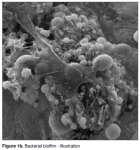
 less likely to develop atopic sensitization or allergic sensitivities (as measured by positive skin-prick tests to common allergens). And, if they have both 'habits', they are even less likely to be allergic to such things as house dust mites, grass, cats, dogs, horses, wool, or airborne fungi. The finding emerges from a longitudinal study which followed the progress of 1,037 persons born in Dunedin, New Zealand in 1972-1973 from childhood into adulthood. However, the researchers found no relationship to these 2 habits to allergic asthma or "hay fever" - a contradictory finding that the researchers don't have an answer for.

"Our findings are consistent with the hygiene theory that early exposure to dirt or germs reduces the risk of developing allergies," said Professor Sears (one of the researchers).  The researchers were testing the idea that the common childhood habits of thumb-sucking and nail-biting would increase microbial exposures, affecting the immune system and reducing the development of allergic reactions also known as atopic sensitization. 31% of the children were frequent thumb suckers or nail biters.

Among all children at 13 years old, 45% showed atopic sensitization, but among those with no habits 49% had allergic sensitization; and those with one oral habit - 40% had allergic sensitization. Among those with both habits, only 31% had allergic sensitization. This trend continued into adulthood, and showed no difference depending on smoking in the household, ownership of cats or dogs; or exposure to house dust mites.

Excerpts of the study from Pediatrics: Thumb-Sucking, Nail-Biting, and Atopic Sensitization, Asthma, and Hay Fever

The hygiene hypothesis suggests that early-life exposure to microbial organisms reduces the risk of developing allergies. Thumb-sucking and nail-biting are common childhood habits that may increase microbial exposures. We tested the hypothesis that children who suck their thumbs or bite their nails have a lower risk of developing atopy, asthma, and hay fever in a population-based birth cohort followed to adulthood. Parents reported children’s thumb-sucking and nail-biting habits when their children were ages 5, 7, 9, and 11 years. Atopic sensitization was defined as a positive skin-prick test (≥2-mm weal) to ≥1 common allergen at 13 and 32 years. 

Thirty-one percent of children were frequent thumb-suckers or nail-biters at ≥1 of the ages. These children had a lower risk of atopic sensitization at age 13 years  and age 32 years. These associations persisted when adjusted for multiple confounding factors. Children who had both habits had a lower risk of atopic sensitization than those who had only 1. No associations were found for nail-biting, thumb-sucking, and asthma or hay fever at either age.

What This Study Adds: Children who sucked their thumbs or bit their nails between ages 5 and 11 years were less likely to have atopic sensitization at age 13. This reduced risk persisted until adulthood. There was no association with asthma or hay fever.

The “hygiene hypothesis” was suggested by Strachan1 to explain why children from larger families and those with older siblings are less likely to develop hay fever. Strahan hypothesized that this could be explained if “allergic diseases were prevented by infection in early childhood transmitted by unhygienic contact with older siblings, or acquired prenatally from a mother infected by contact with her older children.” The hypothesis is supported by evidence showing that children who grow up in large families are at greater risk of coming into contact with more infections....The hygiene hypothesis remains controversial, however, as it is unable to fully explain many associations, including the rise of allergies in “unhygienic” inner-city environments, and why probiotics are ineffective at preventing allergic diseases.3

Thumb-sucking and nail-biting are common oral habits among children, although the reported prevalence varies widely, from <1% to 25%.47 These habits have the potential to increase the exposure to environmental microorganisms, and have been associated with the oral carriage of Enterobacteriaceae, such as Escherichia coli and intestinal parasite infections.812 It seems likely that thumb-sucking and nail-biting would introduce a wide variety of microbes into the body, thus increasing the diversity of the child’s microbiome. If the hygiene hypothesis is correct, it is plausible that this would influence the risk for allergies.... 

Of 1013 children providing data, 317 (31%) had ≥1 oral habit: there was no significant sex difference in prevalence of these habits. Of the 724 children who had skin-prick tests at age 13 years, 328 (45%) showed atopic sensitization. The prevalence of sensitization was lower among children who had an oral habit (38%) compared with those who did not (49%) (P = .009). The lower risk of atopic sensitization was similar for thumb-sucking and nail-biting. Children with only 1 habit were less likely to be atopic (40%) than children with no habit at all (49%), but those with both habits had the lowest prevalence of sensitization (31%) .

An interesting study (published in September 2015) looked at how prevalent biofilms are in the sinuses of people with chronic sinusitis (with or without nasal polyps) as compared to healthy people (without chronic sinusitis). Biofilms are communities of bacteria sticking to one another and coated with a protective slime. The researchers found that the most biofilms were found in people with chronic sinusitis who also had nasal polyps (97.1%) , followed by those with chronic sinusitis without nasal polyps (81.5%), and the least in the control group of healthy patients (56%). They felt that the biofilms contributed to or had a role in chronic sinusitis. But note that the majority of people in all groups had biofilms.

Unfortunately nowhere in the study was there an analysis of the bacteria making up the biofilms. Are the bacteria in the biofilms different in the healthy people versus those with chronic sinusitis? The general assumption is that biofilms are formed from pathogenic (bad) bacteria such as Staphylococcus aureus, but it is known that beneficial bacteria such as Lactobaccillus plantarum and Lactobacillus reuteri can also form biofilms. One study concluded that: "L. reuteri biofilms secreted factors that confer specific health benefits such as immunomodulation and pathogen inhibition." So what was in the biofilms of healthy people (without chronic sinusitis)? Were the biofilms in healthy sinuses made up of protective beneficial bacteria or pathogenic bacteria that were kept in check by other "beneficial" microbes (which can be bacteria, fungi, viruses, etc) in the sinus microbiome?

Biofilms are very hard to eradicate, even with antibiotics. The researchers mentioned that "To date many different modalities have been tested, from Manuka honey to ultrasound and surfactant, but none have been shown to be very efficient." However, they did not mention other bacteria (probiotics) as a treatment possibilty in eradicating biofilms in the sinuses. There has been research looking at using probiotics against biofilms elsewhere in the body (such as dental plaque on teeth).

If biofilms from pathogenic bacteria are so pervasive in chronic sinusitis (81.5% to 97.1%), then it appears that some bacteria such as Lactobacillus sakei somehow predominate over them. I am saying this based on the majority of people writing to me saying that L. sakei treated their chronic sinusitis, as well as the experiences of my own 4 family members (at least 3, perhaps all 4 of us probably had biofilms in our sinuses based on the 81.5% to 97.1% numbers in this research). Something to contemplate. From the journal Acta Oto-Laryngologica:

Bacterial biofilms in chronic rhinosinusitis; distribution and prevalence.

Biofilms were more prevalent in patients with CRSwNP [chronic rhinosinusitis with nasal polyps] compared to both CRSsNP [chronic rhinosinusitis without nasal polyps] and controls [healthy people], and also on the ethmoid bulla compared to the middle turbinate, supporting a biofilm-related pathogenesis of CRSwNP....This study comprised 27 patients with CRSsNP, 34 patients with CRSwNP, and 25 controls.

Chronic rhinosinusitis (CRS) is today understood as a multifaceted group of diseases. The most established differentiation is between CRS with nasal polyps (CRSwNP) and without nasal polyps (CRSsNP)....Patients with CRSwNP have the worst quality-of-life scores, and they have frequent recurrences of their symptoms after surgery.

The pathophysiology of nasal polyps is poorly understood. Bacterial infection, in the form of biofilms, is proposed as a major drive behind the inflammation in CRS. Bacterial biofilms is identified as the agent behind an ever increasing number of chronic infectious diseases, ranging from endocarditis to dental caries. Bacterial biofilms are communities of bacteria in their sessile form, and can be extremely difficult to eradicate with conventional antibiotic therapy.

The total number of patients in the CRS group was 61, 23 females and 38 males, and median age was 40 years....Bacterial biofilms were detected in 97.1% of patients with CRSwNP, 81.5% of patients with CRSsNP, and 56% of controls. Patients with CRSwNP had highly significantly increased prevalence of biofilms compared to controls....The prevalence of biofilms in different anatomical locations within the nasal cavity differed....Biofilms were detected in 79.6% of the samples from the ethmoid bulla, 70.9% of the samples from the uncinated process, and 62.0% of the samples from the middle turbinate.

In this study a significantly increased prevalence of biofilms were found in patients with CRSwNP compared to controls, but also compared to CRSsNP. Indeed only one of the patients with CRSwNP was biofilm negative. This indicates a role for biofilms in the pathogenesis of CRS, but specifically in CRSwNP.

The pathophysiological mechanisms underlying nasal polyps are still poorly understood. Biofilms are shown to be heterogeneous and can be composed of both bacteria and fungi. Staphylococcus Aureus feature prominently in most biofilms found in the sinonasal cavity, being isolated in 50% of the samples. and can possibly facilitate co-colonization with fungi....Bacteria in a biofilm are shown to have up to a 1000-fold increased resistance to antibiotics compared to planktonic bacteria. These features of biofilms make them notoriously hard to eradicate.... In the setting of CRS we have the opportunity of direct local treatment which gives us a greater range of potential treatment options. To date many different modalities have been tested, from Manuka honey to ultrasound and surfactant, but none have been shown to be very efficient....In regards to nasal polyps, further studies are needed to investigate why some patients with biofilms develop nasal polyps while others do not.

Biofilms thrive in moist areas without too much turbulence, conditions found deep in the middle meatus. This may also explain why there were a higher number of biofilm positive CRSwNP patients, as regular nasal polyps originate in the ethmoid....In the opinion of the authors the findings in this article suggest a role for biofilms in CRSwNP.

Bacterial biofilm in a person with chronic sinusitis Credit: Thiago Freire Pinto Bezerra et al,  Braz. j. otorhinolaryngol. (Impr.) vol.75 no.6 São Paulo Nov./Dec. 2009

Of course eating meals prepared at home is healthier! The study results - that people who often consume meals prepared at home are less likely to suffer from type 2 diabetes than those who consume such meals less frequently shouldn't be surprising. The researchers attributed the higher incidence of type 2 diabetes to weight gain in those eating fewer meals prepared at home, but there are other things going on also.

Restaurant and fast food meals tend to have very large portions, frequently with rich sauces, and the meal choices tend to be heavy on fat and salt. The meals can be high in calories, contain many artificial ingredients, and may be low in nutritional quality (and so also not nourishing the beneficial gut microbes that are linked to health). At home you can limit portions, control the food ingredients, and eat only healthy foods (see earlier post on this). From Science Daily:

Enjoying meals prepared at home: Short-cut to avoiding diabetes?

People who often consume meals prepared at home are less likely to suffer from type 2 diabetes than those who consume such meals less frequently, according to  new epidemiological research reported by Qi Sun, of the Harvard T.H. Chan School of Public Heath, Boston, USA and colleagues as part of PLOS Medicine's special issue on Preventing Diabetes.

Internationally, there is an increasing tendency for people to eat out, and this could involve consumption of fast food, for example. Concerns have been raised that such people have a diet that is rich in energy but relatively poor in nutrients -- this could lead to weight gain which is, in turn, associated with an increased risk of type 2 diabetes.

Sun and colleagues employed large prospective data sets in which US health professionals -- both men and women--were followed-up for long periods, with rigorous collection of data on health indicators, including self-reported information on eating habits and occurrence of diabetes. The results were corrected for various known factors that could affect dining habits, including marital status. All in all, the study analyzed 2.1 million years of follow-up data.

The findings indicate that people who reported consuming 5-7 evening meals prepared at home during a week had a 15% lower risk of type 2 diabetes than those who consumed 2 such meals or fewer in a week. A smaller, but still statistically significant, reduction was apparent for those who reported consuming more midday meals prepared at home. Other analyses suggest that less weight gain could partially explain the reported reduction in occurrence of type 2 diabetes in those often eating meals prepared at home.

  Interesting study that supports music instruction for children - that it appears to accelerate brain development in young children, particularly in the areas necessary for general auditory processes such as language, speech and social interaction. Unfortunately music instruction is being cut in many schools, either for budget reasons or because it is perceived as unnecessary. From Developmental Cognitive Neuroscience:

Researchers find that children's brains develop faster with music training

Music instruction appears to accelerate brain development in young children, particularly in the areas of the brain responsible for processing sound, language development, speech perception and reading skills, according to initial results of a five-year study by USC neuroscientists.

These initial study results, published in the journal Developmental Cognitive Neuroscience, provide evidence of the benefits of music education at a time when many schools around the nation have either eliminated or reduced music and arts programs. The study shows music instruction speeds up the maturation of the auditory pathway in the brain and increases its efficiency.

For this longitudinal study, the neuroscientists are monitoring brain development and behavior in a group of 37 children from underprivileged neighborhoods of Los Angeles. Thirteen of the children, at 6 or 7 years old, began to receive music instruction through the Youth Orchestra Los Angeles program at HOLA....The children learn to play instruments, such as the violin, in ensembles and groups, and they practice up to seven hours a week.

The scientists are comparing the budding musicians with peers in two other groups: 11 children in a community soccer program, and 13 children who are not involved in any specific after-school programs. The neuroscientists are using several tools to monitor changes in them as they grow: MRI to monitor changes through brain scans, EEG to track electrical activity in the brains, behavioral testing and other such techniques.

Within two years of the study, the neuroscientists found the auditory systems of children in the music program were maturing faster than in the other children. The fine-tuning of their auditory pathway could accelerate their development of language and reading, as well as other abilities—a potential effect which the scientists are continuing to study. The enhanced maturity reflects an increase in neuroplasticity, a physiological change in the brain in response to its environment—in this case, exposure to music and music instruction.

"The auditory system is stimulated by music," Habibi said. "This system is also engaged in general sound processing that is fundamental to language development, reading skills and successful communication." The auditory system connects our ear to our brain to process sound. When we hear something, our ears receive it in the form of vibrations that it converts into a neural signal. That signal is then sent to the brainstem, up to the thalamus at the center of the brain, and outward to its final destination, the primary auditory cortex, located near the sides of the brain.

A new report authored by dozens of scientists, health practitioners and children's health advocates is highlighting the (growing annually) evidence that many common and widely available chemicals endanger neurological development in fetuses and children of all ages. The chemicals contribute to such health problems as ADHD, autism spectrum disorders, lowered IQ, behavior disorders, and many other problems. Many of the chemicals have hormonal effects (endocrine disruptors) and interfere with normal hormonal activity. The chemicals of highest concern are all around us and are found in most pregnant women, their fetuses, and in growing children. In fact, in all of us.

Especially worrisome chemicals are:  leadmercury; organophosphate pesticides (used in agriculture and home gardens), phthalates (in medicines, plastics, and personal care products), flame retardants known as polybrominated diphenyl ethers (found in upholstered furniture, car seats), air pollutants produced by the combustion of wood and fossil fuels), and polychlorinated biphenyls (once used as coolants and lubricants in electrical equipment, but still pervasive). It is important to note that out of the thousands of chemicals that people are exposed to, that the great majority of chemicals are untested for neurodevelopmental effects.

Especially alarming is that the U.S. Centers for Disease Control found that 90% of pregnant women in the United States have detectable levels of 62 chemicals in their bodies, out of 163 chemicals for which the women were screened. This shows that we are exposed to mixtures of chemicals - not just to one chemical at a time.  Unfortunately the substitutes for problematic chemicals are NO better than the originals, because they tend to be similar chemically. For example, the substitutes for BPA are just as bad, if not worse, than BPA (bisphenol A). And remember, we are exposed to mixtures of chemicals - not just to one chemical at a time.

The report criticizes current regulatory lapses that allow chemicals to be introduced into people's lives with little or no review of their effects on fetal and child health. "For most chemicals, we have no idea what they're doing to children's neurodevelopment," Professor Schantz (one of the signers of the report) said. "They just haven't been studied." So why aren't policymakers doing something? Why is industry dictating what we're exposed to? Why are chemicals innocent until proven guilty, and even then they're allowed to be used? Who is looking out for the ordinary person, and especially developing children?

From the journal Environmental Health Perspectives: Project TENDR: Targeting Environmental Neuro-Developmental Risks. The TENDR Consensus Statement

Children in America today are at an unacceptably high risk of developing neurodevelopmental disorders that affect the brain and nervous system including autism, attention deficit hyperactivity disorder, intellectual disabilities, and other learning and behavioral disabilities. These are complex disorders with multiple causes—genetic, social, and environmental. The contribution of toxic chemicals to these disorders can be prevented. 

Leading scientific and medical experts, along with children’s health advocates, came together in 2015 under the auspices of Project TENDR: Targeting Environmental Neuro-Developmental Risks to issue a call to action to reduce widespread exposures to chemicals that interfere with fetal and children’s brain development. Based on the available scientific evidence, the TENDR authors have identified prime examples of toxic chemicals and pollutants that increase children’s risks for neurodevelopmental disorders. These include chemicals that are used extensively in consumer products and that have become widespread in the environment. Some are chemicals to which children and pregnant women are regularly exposed, and they are detected in the bodies of virtually all Americans in national surveys conducted by the U.S. Centers for Disease Control and Prevention. The vast majority of chemicals in industrial and consumer products undergo almost no testing for developmental neurotoxicity or other health effects.

Based on these findings, we assert that the current system in the United States for evaluating scientific evidence and making health-based decisions about environmental chemicals is fundamentally broken. To help reduce the unacceptably high prevalence of neurodevelopmental disorders in our children, we must eliminate or significantly reduce exposures to chemicals that contribute to these conditions. We must adopt a new framework for assessing chemicals that have the potential to disrupt brain development and prevent the use of those that may pose a risk. This consensus statement lays the foundation for developing recommendations to monitor, assess, and reduce exposures to neurotoxic chemicals. 

The TENDR Consensus Statement is a call to action to reduce exposures to toxic chemicals that can contribute to the prevalence of neurodevelopmental disabilities in America’s children. The TENDR authors agree that widespread exposures to toxic chemicals in our air, water, food, soil, and consumer products can increase the risks for cognitive, behavioral, or social impairment, as well as specific neurodevelopmental disorders such as autism and attention deficit hyperactivity disorder (ADHD) (Di Renzo et al. 2015; Gore et al. 2015; Lanphear 2015; Council on Environmental Health 2011). This preventable threat results from a failure of our industrial and consumer markets and regulatory systems to protect the developing brain from toxic chemicals. To lower children’s risks for developing neurodevelopmental disorders, policies and actions are urgently needed to eliminate or significantly reduce exposures to these chemicals.

We are witnessing an alarming increase in learning and behavioral problems in children. Parents report that 1 in 6 children in the United States, 17% more than a decade ago, have a developmental disability, including learning disabilities, ADHD, autism, and other developmental delays (Boyle et al. 2011). As of 2012, 1 in 10 (> 5.9 million) children in the United States are estimated to have ADHD (Bloom et al. 2013). As of 2014, 1 in 68 children in the United States has an autism spectrum disorder (based on 2010 reporting data) (CDC 2014).

Many toxic chemicals can interfere with healthy brain development, some at extremely low levels of exposure. Research in the neurosciences has identified “critical windows of vulnerability” during embryonic and fetal development, infancy, early childhood and adolescence (Lanphear 2015; Lyall et al. 2014; Rice and Barone 2000). During these windows of development, toxic chemical exposures may cause lasting harm to the brain that interferes with a child’s ability to reach his or her full potential.

The developing fetus is continuously exposed to a mixture of environmental chemicals (Mitro et al. 2015). A 2011 analysis of the U.S. Centers for Disease Control and Prevention’s (CDC) biomonitoring data found that 90% of pregnant women in the United States have detectable levels of 62 chemicals in their bodies, out of 163 chemicals for which the women were screened (Woodruff et al. 2011). Among the chemicals found in the vast majority of pregnant women are PBDEs, polycyclic aromatic hydrocarbons (PAHS), phthalates, perfluorinated compounds, polychlorinated biphenyls (PCBs), perchlorate, lead and mercury (Woodruff et al. 2011). Many of these chemicals can cross the placenta during pregnancy and are routinely detected in cord blood or other fetal tissues.

The following list provides prime examples of toxic chemicals that can contribute to learning, behavioral, or intellectual impairment, as well as specific neurodevelopmental disorders such as ADHD or autism spectrum disorder: Organophosphate (OP) pesticides, PBDE flame retardants, combustion-related air pollutants, which generally include PAHs, nitrogen dioxide and particulate matter, and other air pollutants for which nitrogen dioxide and particulate matter are markers, lead, mercuryPCBs .

The United States has restricted some of the production, use and environmental releases of these particular chemicals, but those measures have tended to be too little and too late. We face a crisis from both legacy and ongoing exposures to toxic chemicals.....The examples of developmental neurotoxic chemicals that we list here likely represent the tip of the iceberg....Only a minority of chemicals has been evaluated for neurotoxic effects in adults. Even fewer have been evaluated for potential effects on brain development in children (Grandjean and Landrigan 2006, 2014). Further, toxicological studies and regulatory evaluation seldom address combined effects of chemical mixtures, despite evidence that all people are exposed to dozens of chemicals at any given time.

Some chemicals, like those that disrupt the endocrine system, present a concern because they interfere with the activity of endogenous hormones that are essential for healthy brain development. Endocrine-disrupting chemicals (EDCs) include many pesticides, flame retardants, fuels, and plasticizers. One class of EDCs that is ubiquitous in consumer products are the phthalates. These are an emerging concern for interference with brain development and therefore demand attention.

Under our current system, when a toxic chemical or category of chemicals is finally removed from the market, chemical manufacturers often substitute similar chemicals that may pose similar concerns or be virtually untested for toxicity. This practice can result in “regrettable substitution” whereby the cycle of exposures and adverse effects starts all over again. The following list provides examples of this cycle: When the federal government banned some uses of OP pesticides, manufacturers responded by expanding the use of neonicotinoid and pyrethroid pesticides. Evidence is emerging that these widely used classes of pesticides pose a threat to the developing brain (Kara et al. 2015; Richardson et al. 2015; Shelton et al. 2014). 

When the U.S. Government reached a voluntary agreement with flame retardant manufacturers to stop making PBDEs, the manufacturers substituted other halogenated and organophosphate flame retardant chemicals. Many of these replacement flame retardants are similar in structure to other neurotoxic chemicals but have not undergone adequate assessment of their effects on developing brains. When the federal government banned some phthalates in children’s products, the chemical industry responded by replacing the banned chemicals with structurally similar new phthalates. These replacements are now under investigation for disrupting the endocrine system.