Skip to content

A new study has summarized what we know about fungi that live in and on babies - and yes, we all have fungi both on and within us. It's called the mycobiome. In healthy individuals all the microbes (bacteria, viruses, fungi, etc) live in balanced microbial communities, but the communities can become "out of whack" (dysbiosis) for various reasons, and microbes that formerly co-existed peacefully can multiply and become problematic. Or other pathogenic microbes can enter the community, and the person becomes ill.

In healthy adults, approximately 0.1% of the microbes in the adult intestine are fungi, from approximately 60 unique species. Most species live peacefully in the body, and some fungi even have health benefits (e.g., Saccharomyces boulardii prevents gastrointestinal disease). Some fungi that many view as no good and involved with diseases (e.g., Candida and Aspergillus) are also found normally in healthy people. Studies show that normally infants also have fungi. Some fungi that live in the baby's gut (thus detected in fecal samples) are Candida (including C. albicans), Saccharomyces, and Cladosporium. The researchers (from the Univ. of Minnesota) point out that the study of fungi in babies has been neglected and much more research needs to be done.

Whether an infant is born vaginally or through cesarean delivery (C-section) affects the composition of the baby's bacterial communities over the first 6 months of life. And similarly, it looks like when the baby passes through the birth canal, the baby is exposed to the mother's mycobiota (fungi), and then these colonize in the infant's gut. Babies born by C-section have some differences in their fungi, such as being colonized by the mother's skin fungi (such as Malassezia fungi). After birth, a parent kissing and touching the baby (skin to skin contact) also transmits microbes, including fungi, to the baby.

Whether a baby drinks breast milk or formula strongly affects the infant's bacteria within the GI tract. For example, breast-fed infants have more Bifidobacteria and Labctobacilli in their gut compared to formula-fed infants. One study found about 700 species of bacteria in breast milk. Thus, scientists think that human breast milk also influences the infant gut mycobiota (fungi), although this research still needs to be done.

Whether a baby is born prematurely or at term (gestational age) is important. For infants born prematurely, intestinal fungi can cause big problems, such as an overgrowth in the gut. For example, 10% of premature babies get invasive, systemic Candidiasis, and about 20% die. Some factors leading to this are: a naïve immune system, bacterial communities out of whack (dysbiosis) due to antibiotic exposure, and use of parenteral nutrition (because this doesn't contain all the microbes from the mother that are in breast milk). In premature infants, beneficial fungi such as S. boulardii, may help to regulate the growth of opportunistic fungal colonizers such as Candida.

it is clear that whether the baby received antibiotics is important. The bacterial community of infants is altered by exposure to antibiotics in both term and preterm infants. For example, in a lengthy study over the first 3 years of life, infants receiving multiple courses of antibiotics had bacterial community changes following antibiotics and their gut bacterial microbiome became less diverse (fewer species). Although most commonly used antibiotics do not directly act on fungi, anti-bacterial antibiotic exposure is associated with alterations to the mycobiota (fungi) -  such as increased rates of fungal colonization, fungal overgrowth, and changes in the fungal community. For ex., premature infants exposed to cephalosporin antibiotics have an increased risk for invasive Candidiasis (a fungal overgrowth).

Out of whack (dysbiotic) microbial communities, incuding fungi, are found in IBD (intestinal bowel diseases) in children. They have more of some fungi (e.g. Pichia jadinii and Candida parapsilosis) and less of Cladosporium cladosporiodes, and an overall decrease in fungal diversity in the gut, as compared to healthy children.

From BMC Medicine: Infant fungal communities: current knowledge and research opportunities

The microbes colonizing the infant gastrointestinal tract have been implicated in later-life disease states such as allergies and obesity. Recently, the medical research community has begun to realize that very early colonization events may be most impactful on future health, with the presence of key taxa required for proper immune and metabolic development. However, most studies to date have focused on bacterial colonization events and have left out fungi, a clinically important sub-population of the microbiota. A number of recent findings indicate the importance of host-associated fungi (the mycobiota) in adult and infant disease states, including acute infections, allergies, and metabolism, making characterization of early human mycobiota an important frontier of medical research. This review summarizes the current state of knowledge with a focus on factors influencing infant mycobiota development and associations between early fungal exposures and health outcomes. We also propose next steps for infant fungal mycobiome research....

A new study found differences in gut microbes between active women (they exercised at least the recommended amount) and those that are sedentary. When the gut bacteria were analyzed with modern tests (genetic sequencing) the active women had more of the health promoting beneficial bacteria such as Faecalibacterium prausnitzii, Roseburia hominis, and Akkermansia muciniphila than the sedentary women. The sedentary women also had some bacterial species not seen in the active women. The researchers said that exercise "modifies the composition of gut microbiota" (the gut microbes) in a way beneficial for health.

And what is the recommended minimal amount of exercise? The World Health Organization recommends at least 3 days of exercise per week for 30 minutes at a moderate intensity. Note that exercise can mean doing exercises, but it can also include walking briskly, intense housework (scrubbing, vacuuming with lots of bending, etc.), gardening (digging, raking, etc), or shoveling snow, etc. In this study the group of active women had at least 3 hours of physical exercise per week. Note that a sedentary lifestyle is associated with a high incidence of chronic diseases such as cardiovascular disease, cancer and diabetes, while physical exercise or activity has metabolic and immune health benefits (prevents disease).

But...reading the full study, the research also showed that the active group ate more fruits and vegetables - which we know has an effect on the gut microbiome and feeds beneficial bacteria. Although the diets of the 2 groups of women were similar in total carbohydrates, protein and fat content eaten, the active women ate more fruits, vegetables, and fiber, and the sedentary group ate more processed meat. So it looks like both exercise and a good amount of fruits and vegetables may be important for nurturing beneficial bacteria. By the way, the 3 species of beneficial bacteria mentioned currently are not found in any probiotic supplements on the market. (Earlier posts on the beneficial F. prausnitzii and Akkermansia muciniphila). From C. Bressa et al research article in PLoS ONE:

Differences in gut microbiota profile between women with active lifestyle and sedentary women

Physical exercise is a tool to prevent and treat some of the chronic diseases affecting the world’s population. A mechanism through which exercise could exert beneficial effects in the body is by provoking alterations to the gut microbiota, an environmental factor that in recent years has been associated with numerous chronic diseases. Here we show that physical exercise performed by women to at least the degree recommended by the World Health Organization can modify the composition of gut microbiota. Using high-throughput sequencing of the 16s rRNA gene, eleven genera were found to be significantly different between active and sedentary women. Quantitative PCR analysis revealed higher abundance of health-promoting bacterial species in active women, including Faecalibacterium prausnitzii, Roseburia hominis and Akkermansia muciniphila. Moreover, body fat percentage, muscular mass and physical activity significantly correlated with several bacterial populations. In summary, we provide the first demonstration of interdependence between some bacterial genera and sedentary behavior parameters, and show that not only does the dose and type of exercise influence the composition of gut microbiota, but also the breaking of sedentary behavior. ...continue reading "Gut Microbe Differences Between Active and Sedentary Women"

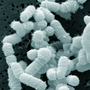
After writing about Lactobacillus sakei in the sinuses for several years (present in healthy sinuses, absent or less in those with chronic sinusitis, and also a treatment for chronic sinusitis), I wondered whether L. sakei is found anywhere else in the body. Today I read a study (conducted in Japan) about gut microbes and strokes and there it was - the presence of L. sakei in the gut.

Specifically, a study found that people who have ischemic strokes tend to have lower amounts ("depletion") of L. sakei in the gut than healthy people, even though it was detected in 80% of both groups.

The study found that in people with ischemic strokes there was evidence for the gut microbes being out of whack (dysbiosis), as well as more inflammation, and more of certain bacteria species (Atopobium cluster and Lactobacillus ruminis), and depletion of L. sakei bacteria.

The researchers took samples of stool (fecal samples) from each person of both groups (ischemic stroke group and healthy group) and analyzed the stool with modern tests (genetic sequencing) to see whether 22 groups of bacteria were in it. (Note that there are normally hundreds of species of bacteria living in a healthy person's gut, as well as viruses, fungi, etc.).

So once again it looks like L. sakei may be beneficial bacteria, even in the gut. The researchers were careful to point out that they couldn't say that certain bacteria caused the strokes - just that there was an association.

And what diet is associated with lower levels of inflammation in the body? Once again - a diet with lots of fruits, vegetables, whole grains, nuts, seeds, and legumes (think Mediterranean style diet). You want to feed the beneficial bacteria in the gut.

Excerpts from a research article by Yamashiro et al in PLoS One: Gut dysbiosis is associated with metabolism and systemic inflammation in patients with ischemic stroke

The role of metabolic diseases in ischemic stroke has become a primary concern in both research and clinical practice. Increasing evidence suggests that dysbiosis is associated with metabolic diseases. The aim of this study was to investigate whether the gut microbiota, as well as concentrations of organic acids, the major products of dietary fiber fermentation by the gut microbiota, are altered in patients with ischemic stroke, and to examine the association between these changes and host metabolism and inflammation.

We analyzed the composition of the fecal gut microbiota and the concentrations of fecal organic acids in 41 ischemic stroke patients and 40 control subjects via 16S and 23S rRNA-targeted quantitative reverse transcription (qRT)-PCR and high-performance liquid chromatography analyses..... Although only the bacterial counts of Lactobacillus ruminis were significantly higher in stroke patients compared to controls, multivariable analysis showed that ischemic stroke was independently associated with increased bacterial counts of Atopobium cluster and Lactobacillus ruminis, and decreased numbers of Lactobacillus sakei subgroup, independent of age, hypertension, and type 2 diabetes....Together, our findings suggest that gut dysbiosis in patients with ischemic stroke is associated with host metabolism and inflammation.   ...continue reading "Gut Bacteria Associated With Strokes"

Interesting idea - that perhaps our community of gut microbes being out of whack (dysbiosis) leads to hypertension. This study was done in both humans and mice - with an analysis of bacteria in both hypertensive individuals and pre-hypertensives, and also healthy individuals (the controls). Then the microbes from 2 hypertensive individuals were transplanted into mice (fecal microbiota transplants). And lo and behold - the mice became hypertensive with an alteration of their gut microbes. This is amazing!

The study showed that transplanting microbes from hypertensives to non-hypertensives caused an elevation in blood pressure in the formerly healthy group. This shows the direct influence of gut microbes on blood pressure. The bacteria found in both the pre-hypertensives and hypertensives (especially an overgrowth of Prevotella and Klebsiella bacteria) are those linked to inflammation. And what kind of diet is linked to that bacteria? A high fat diet. Yes, the Western diet with lots of fat and highly processed foods.

The researchers talked about other research also showing Prevotella being associated not only with hypertension, but also other diseases (e.g., periodontal diseases and rheumatoid arthritis). On the other hand, Faecalibacterium, Oscillibacter, Roseburia, Bifidobacterium, Coprococcus, and Butyrivibrio, which were "enriched" in healthy controls, were lower in pre-hypertensive and hypertensive persons. In the past I have posted about a "special" bacteria that is even called  a "keystone" gut bacteria - Faecalibacterium prausnitziithat is linked to health and is low or absent in the gut in a number of diseases ((here and here). It is not available in a supplement at this time (because it dies within a few minutes upon exposure to oxygen), but diet influences it. A high animal meat, high animal fat, high sugar, highly processed foods, and low fiber diet (the typical Western diet) lowers F. prausnitzii numbers, while a high-fiber, low meat diet increases F. prausnitzii numbers.

What you can do: Feed the beneficial gut microbes by increasing the amount of fruits, vegetables, whole grains, seeds, nuts that you eat. And cut back on the greasy, high fat processed and fast foods.

The following excerpt is misleading - for example, it ignores the first part of the actual study which looked at the gut bacteria of pre-hypertensives, hypertensive, and healthy people. Then gut bacteria from hypertensive people were transplanted into healthy mice, and gut bacteria from healthy people were transplanted into hypertensive mice. Also, it wasn't rats, but mice used in the study. It goes to show why it's important to look at original studies - not just believe articles out there blindly. [See original study.] From Science Daily: Unhealthy gut microbes a cause of hypertension, researchers find

Researchers have found that the microorganisms residing in the intestines (microbiota) play a role in the development of high blood pressure in rats mice....Scientists studied two sets of rats mice, one group with high blood pressure ("hypertensive") and one with normal blood pressure ("normal").... All animals were then given antibiotics for 10 days to reduce their natural microbiota. After the course of antibiotics, the researchers transplanted hypertensive microbiota to normal blood pressure rats mice and normal microbiota to the hypertensive group. 

The researchers found that the group treated with hypertensive microbiota developed elevated blood pressure. A more surprising result is that the rats mice treated with normal microbiota did not have a significant drop in blood pressure, although readings did decrease slightly. This finding is "further evidence for the continued study of the microbiota in the development of hypertension in humans and supports a potential role for probiotics as treatment for hypertension," wrote the researchers. "Studies showing that supplementing the diet with probiotics (beneficial microorganisms found in the gut) can have modest effects on blood pressure, especially in hypertensive models."

NOTE that the actual study said in its CONCLUSIONS:  "Taken together, we have described clearly the disordered profiles of gut microbiota and microbial products in human patients with pre-hypertension and hypertension, established the relationship between gut dysbiosis and hypertension, and provided important evidence for the novel role of gut microbiota dysbiosis as a key factor for blood pressure changes. Our findings point towards a new strategy aimed at preventing the development of hypertension and reducing cardiovascular risks through restoring the homeostasis of gut microbiota, by improving diet and lifestyle or early intervening with drugs or probiotics."

What things in our environment have an effect on the microbes living within us? We now know that gut microbes are important for our health in many ways, and that thousands of species of bacteria, as well as viruses, fungi, and other microbes normally live in a healthy person's gut. We refer to these microbes as the human microbiota or human microbiome. When the community of gut microbes is thrown out of whack (dysbiosis) there can be a number of negative health effects, including diseases. Researchers are just learning about all the microbes within us and their importance in health and disease. [See all posts on the human microbiome.]

Past posts have discussed such things as antibiotics, emulsifiers, different foods and diets, heartburn drugs, etc. having an effect on the human microbiome, but what else? A recent study from China reviewed some environmental pollutants and their effects on gut microbiota - as shown in both human and animal studies. They reviewed studies on antibiotics, heavy metals (arsenic, cadmium, lead), persistant organic pollutants or POPs (organochlorine pesticides, polychlorinated biphenyls (PCBs), polybrominated diphenyl ethers, and polycyclic aromatic hydrocarbons or PAHs), pesticides (permethrin, chlorpyrifos, pentachlorophenol, epoxiconazole and carbendazim, imazalil), emulsifiers, nanoparticles (e.g., silver nanoparticles), and artificial sweeteners. They found that all these environmental pollutants had effects on gut microbes - with some effects lasting for years. Their conclusion: gut microbes are very sensitive to drugs, diet, and environmental pollutants. By the way, notice that popular food ingredients such as emulsifiers and artificial sweeteners were considered "environmental pollutants" by the researchers.

Excerpts from Environmental Pollution: Effects of environmental pollutants on gut microbiota

Environmental pollutants have become an increasingly common health hazard in the last several decades. Recently, a number of studies have demonstrated the profound relationship between gut microbiota and our health. Gut microbiota are very sensitive to drugs, diet, and even environmental pollutants. In this review, we discuss the possible effects of environmental pollutants including antibiotics, heavy metals, persistent organic pollutants, pesticides, nanomaterials, and food additives on gut microbiota and their subsequent effects on health. We emphasize that gut microbiota are also essential for the toxicity evaluation of environmental pollution. In the future, more studies should focus on the relationship between environmental pollution, gut microbiota, and human health.

Thousands of species are found in the gut microbiome, and the majority of these species belong to six bacterial phyla: Firmicutes, Bacteroidetes, Actinobacteria, Proteobacteria, Fusobacteria, and Verrucomicrobia (Eckburg et al., 2005). Gut microbiota are highly dynamic and have substantial interindividual and intraindividual variation....The gut microbiota are very essential for host health. They participate in the regulation of many physiological functions. The gut microbiota reside in our intestinal mucus layer and even participate in shaping the mucus layer (Jakobsson et al., 2015). They help us to digest food (such as fiber); synthesize vitamins and amino acids (Spanogiannopoulos et al., 2016); play very important roles in energy metabolism and storage, immune system modulation, growth, and neurodevelopment; and can even regulate our behavior.... The occurrence of many diseases is correlated with altered gut microbiome composition (Lange et al., 2016). Gut microbiota dysbiosis is considered to be a potential cause of obesity (Cani et al., 2007; Fei and Zhao, 2013). However, gut microbiota are very sensitive to drugs, diet, and environmental pollutants.

Although most environmental pollutants do not directly target gut microbiota, some pollutants can enter the body and interact with the gut microbiota through different pathways. A number of previous studies have shown that exposure to environmental pollutants can alter the composition of the gut microbiome, leading to disorders of energy metabolism, nutrient absorption, and immune system function or the production of other toxic symptoms (Jin et al., 2015c; Zhang et al., 2015b). In the present review, we conclude that different kinds of environmental pollutants can induce gut microbiota dysbiosis and have multiple potential adverse effects on animal health

Heavy metals in the environment have become a severe health risk in recent years (Liu et al., 2016a). As a common form of environmental pollution, heavy metals are associated with a wide range of toxic effects, including carcinogenesis, oxidative stress, and DNA damage, and effects on the immune system..... Recently, several studies have stated that heavy metal exposure could also lead to gut microbiota dysbiosis, indicating that study of gut microbiota provides a new approach to analyze the mechanisms of heavy metal toxicity

Immune system function is tightly coupled to our gut microbiome. Gut microbiota and their metabolites can interact with both the innate immune system and the adaptive immune system (Honda and Littman, 2016; Thaiss et al., 2016).... Alterations in the gut microbiome can disrupt the balance between the host immune system and gut microbiota, induce immune responses, and even trigger some immunological diseases. Furthermore, immune system imbalance may influence the microbiota metabolites. For example, trimethylamine, which is absorbed from food by gut microbiota, can induce atherosclerosis (Chistiakov et al., 2015).

For years it has been known that most children with autism spectrum disorder (ASD) have all sorts of gastrointestinal (GI) problems (e.g., constipation, diarrhea, stomach pain, food intolerance), and the more severe the autism, the more severe the GI problems. Recent studies suggested that a major factor in this are abnormal gut bacteria, with the gut microbial community out of whack (dysbiosis). Previous studies looking at the gut microbiome of children with autism have shown lower diversity and lower amounts (abundances) of certain bacteria in children with autism compared to neurologically normal (neurotypical) children.

A recent study of children with autism spectrum disorder found that giving the children a fecal microbiota transplant (FMT) led to significant and lasting improvements in both gastrointestinal (GI) symptoms and autism-related behaviors and symptoms. A fecal microbiota transplant (FMT) is a transplant of fecal matter from a healthy donor to the recipient. A fecal microbial transplant contains approximately a thousand bacterial species that live in a healthy gut, as well as other microbes such as viruses and fungi. FMTs have so far been an amazingly successful treatment for recurrent Clostridium difficile infections, and are now being looked at as promising treatments of chronic inflammatory diseases such as inflammatory bowel disease.

The researchers were surprised to see an 80% improvement in gastrointestinal symptoms, especially abdominal pain, indigestion, diarrhea, and constipation. They also saw about a 25% improvement in autism related behaviors and symptoms which persisted for 8 weeks after treatment stopped, which is when the study ended. One measurement of adaptive behaviors (such as communication, daily living skills, and socialization) found that the average developmental age increased by 1.4 years after treatment. The researchers also found that there was a "rebalancing" of the gut microbes following treatment. They found evidence of "successful partial engraftment of donor microbiota and beneficial changes in the gut environment" - meaning they could see that donor microbes were living in the gut. Also, overall bacterial diversity increased (which is good) and the abundance of certain bacteria increased (including Bifidobacterium, Prevotella, and Desulfovibrio), and these changes persisted until the end of the study.

The researchers caution that this was a small trial, that there could be placebo effects, and so the results should be "cautiously interpreted and viewed as preliminary." But nonetheless, the results are exciting. Really exciting. From Science Daily:

Autism symptoms improve after fecal transplant, small study finds

Children with autism may benefit from fecal transplants -- a method of introducing donated healthy microbes into people with gastrointestinal disease to rebalance the gut, a new study has found. Behavioral symptoms of autism and gastrointestinal distress often go hand-in-hand, and both improved when a small group of children with the disorder underwent fecal transplant and subsequent treatment. In the study of 18 children with autism and moderate to severe gastrointestinal problems, parents and doctors said they saw positive changes that lasted at least eight weeks after the treatment. Children without autism were included for comparison of bacterial and viral gut composition prior to the study.

Previous research has established that children with autism typically have fewer types of some important bacteria in their guts and less bacterial diversity overall -- a difference that held true in this study. That could be because many of them are prescribed a lot of antibiotics in the first three years of life, the research team wrote in the study.

Parents of the children not only reported a decrease in gut woes including diarrhea and stomach pain in the eight weeks following the end of treatment: They also said they saw significant changes for the better when it came to behavioral autism symptoms in their sons and daughters, who ranged from 7 to 16 years old....One of those tools showed the average developmental age increased by 1.4 years after treatment. 

Researchers also were able to document a rebalancing of the gut following treatment. At the end of the study, the bacterial diversity in the children with autism was indistinguishable from their healthy peers. The study also included a unique viral analysis by Ohio State scientists, made possible because of previous work in the world's oceans. Gregory, who is particularly interested in the interplay between viruses and bacteria, used genetic testing to examine the viral diversity in the guts of the treated children. It rebounded quickly, and became more similar to the donor's microbiome. "Those donor viruses seemed to help," she said.

Fecal transplantation is done by processing donor feces and screening it for disease-causing viruses and bacteria before introducing it into another person's gastrointestinal tract. In this study, the researchers used a method called microbiota transfer therapy, which started with the children receiving a two-week course of antibiotics to wipe out much of their existing gut flora. Then, doctors gave them an initial high-dose fecal transplant in liquid form. In the seven to eight weeks that followed, the children drank smoothies blended with a lower-dose powder[Original study.]

What exactly are the differences between people with chronic sinusitis and those who are healthy and don't get sinusitis? I've written many times about the Abreu et al 2012 study that found that not only do chronic sinusitis sufferers lack L. sakei, they have too much of Corynebacterium tuberculostearicum (normally a harmless skin bacteria), and they also don't have the bacteria diversity in their sinuses that healthy people without sinusitis have.

In other words, the sinus microbiome (microbial community) is out of whack (dysbiosis). A number of studies found that there is a depletion of some bacterial species, and an increase in "abundance" of other species in those with chronic sinusitis.

Now a new analysis of 11 recent studies comparing people with chronic sinusitis to healthy people adds some additional information. Once again a conclusion was that those with sinusitis had "dysbiosis" (microbial communities out of whack) in their sinus microbiomes when compared to healthy people. And that an increased "abundance" of members of the genus Corynebacterium in the sinuses was associated with chronic sinusitis (studies so far point to C. tuberculostearicum and C. accolens). Nothing new there...

But what was new was that they found that bacteria of the genus Burkholderia and Propionibacterium seem to be "gatekeepers", whose presence may be important in maintaining a stable and healthy bacterial community in the sinuses. And that in chronic sinusitis the bacterial network of healthy communities is "fragmented". In other words, when a person is healthy, the community of microbes in the sinuses may provide a protective effect, and if the gatekeepers are removed (e.g., during illnesses or after taking antibiotics), then a "cycle of dysbiosis and inflammation" may begin.

PLEASE NOTE: Genus is a taxonomic category ranking used in biological classification that is below a family and above a species level. For example, Lactobacillus is the genus and sakei is the species. Also, the researchers discussed "gatekeepers" as being important for sinus health, while Susan Lynch discusses the importance of "keystone species" for sinus health.

OK... so which species of Burkholderia and Propionibacterium bacteria are found in the healthy microbiome? Unfortunately that was not answered in this study. And of course this needs to be tested further to see if the addition of the missing species of Burkholderia and Propionibacterium bacteria to the sinus microbiome will treat chronic sinusitis. Or perhaps other bacteria such as L. sakei and someother still unknown bacteria also need to be added to the mix.

Both Burkholderia and Propionibacterium have many species, but I have not seen any in probiotics. Species of Propionibacteria can be found all over the body and are generally nonpathogenic. However, P. acnes can cause the common skin condition acne as well as other infections.

One species - Propionibacterium freudenreichii (or P. shermanii)  - is found in Swiss type cheeses such as Emmental, Jarlsberg, and Leerdammer. Propionibacteria species are commonly found in milk and dairy products, though they have also been extracted from soil. There are many Burkholderia species, with a number of them causing illness (e.g., B. mallei and B. pseudomallei), but also beneficial species, such as those involved with plant growth and healthBurkholderia species are found all over, in the soil, in plants, soil, water (including marine water), rhizosphere, animals and humans. At this point it is unclear to me which are the species found in healthy sinuses.

But it is clear that while L. sakei works to treat chronic sinusitis in many people, the fact that L. sakei typically has to be used after each illness (cold, sore throat, etc,) means that the sinus microbiome may still be missing microbial species or that there is still some sort of "imbalance" (even though the person may feel totally healthy). The researchers noted that a variety of fungi and viruses are also part of a normal sinus microbiome, but they weren't discussed in the article. As you can see, much is still unknown. Stay tuned..,..

This was a very technical article - thus not easy to read. Keep in mind that the information about the conclusions about the bacteria species in the sinuses was from studies that used modern genetic sequencing data (16S rRNA sequence data) to determine what bacteria are in the sinuses. (These are called "culture independent technologies" and much, much better than using cultures in determining species of bacteria.) This way they could analyze differences in "sinonasal bacterial community composition" and see differences between healthy people and persons with CRS (chronic rhinosinusitis).

Excerpts from Environmental Microbiology: Bacterial community collapse: a meta-analysis of the sinonasal microbiota in chronic rhinosinusitis

Chronic rhinosinusitis (CRS) is a common, debilitating condition characterized by long-term inflammation of the nasal cavity and paranasal sinuses. The role of the sinonasal bacteria in CRS is unclear. We conducted a meta-analysis combining and reanalysing published bacterial 16S rRNA sequence data to explore differences in sinonasal bacterial community composition and predicted function between healthy and CRS affected subjects. The results identify the most abundant bacteria across all subjects as Staphylococcus, Propionibacterium, Corynebacterium, Streptococcus and an unclassified lineage of Actinobacteria.

The meta-analysis results suggest that the bacterial community associated with CRS patients is dysbiotic and ecological networks fostering healthy communities are fragmented. Increased dispersion of bacterial communities, significantly lower bacterial diversity, and increased abundance of members of the genus Corynebacterium are associated with CRS. Increased relative abundance and diversity of other members belonging to the phylum Actinobacteria and members from the genera Propionibacterium differentiated healthy sinuses from those that were chronically inflamed. Removal of Burkholderia and Propionibacterium phylotypes from the healthy community dataset was correlated with a significant increase in network fragmentation. This meta-analysis highlights the potential importance of the genera Burkholderia and Propionibacterium as gatekeepers, whose presence may be important in maintaining a stable sinonasal bacterial community.

The high density and diversity of host-associated microbial communities present in different body sites supports a near infinite number of potential host to microbe, and microbe to microbe interactions. A stable network of microbial interactions, established through processes such as niche competition, nutrient cycling, immune evasion, and biofilm formation help maintain homeostasis during health (Walter and Ley, 2011; Grice et al., 2009). Taxa that hold together the bacterial community by interacting with different parts of the network can be considered “gatekeepers” (sensu Freeman, 1980; Widder et al., 2014). During health, a consortium of microbes may provide a protective effect, and a breakdown in these networks due to the removal of gatekeepers may begin a self-perpetuating cycle of dysbiosis and inflammation (Vujkovic-Cvijin et al., 2013; Widder et al., 2014; Byrd and Segre, 2016).

The genus-level phylotype Corynebacterium was again associated with CRS bacterial communities, and Burkholderia was associated with healthy subjects.

In contrast to the variety of Actinobacteria and Betaproteobacteria phylotypes differentiating the healthy sinonasal bacterial communities, only one phylotype (Corynebacterium) was consistently associated with those individuals that were chronically inflamed. The significance of specific members of the genus Corynebacterium in CRS microbial communities is supported by findings in two previous studies (Abreu et al., 2012; Aurora et al., 2013). The relative abundance of C. tuberculostearicum and C. accolens was significantly higher in subjects with CRS in two recent 16S rRNA studies (Abreu et al., 2012 and Aurora et al., 2013, respectively). 

 The research finding of dogs having elevated levels of the endocrine disruptor bisphenol A (BPA) from eating canned food mirrors what is happening to humans - eating canned food raises BPA levels in a person. The study also found that elevated BPA levels resulted in changes in the gut microbiome (the community of microbes living in the gut). Specifically, they found the abundance of a number of bacteria species increased or decreased depending on BPA levels in the dogs. This is not good.

This is of concern because BPA  is linked to a variety of health problems. [See all posts.] So it's best to minimize exposure to BPA, BPS, and other hormone disrupting chemicals, and also "BPA-free" products (which usually contain BPS). The BPA is in the lining of the cans used in canned food, and this leaches into the food. Unfortunately, dog food cans thought to be BPA-free in the study also contained BPA, which then leached into the dog food.  From Futurity:

Dogs have 3X more BPA after eating canned food

Researchers saw a three-fold increase in BPA levels in dogs who ate canned dog food for two weeks. They also saw changes in the dogs’ gut microbesBisphenol A (BPA) is a widely used industrial chemical found in many household items, including resins used to line metal storage containers, such as food cans. The chemical can disrupt hormones and is linked to a range of health problems. “Bisphenol A is a prevalent endocrine-disrupting chemical found in canned foods and beverages,” says Cheryl Rosenfeld, an associate professor of biomedical sciences in the University of Missouri College of Veterinary Medicine....

Dog owners volunteered their healthy pets for the study. Blood and fecal samples were collected prior to the dogs being placed on one of two commonly used, commercial canned food diets for two weeks; one diet was presumed to be BPA-free. Robert Backus, an associate professor in the veterinary medicine and surgery in the College of Veterinary Medicine, and other researchers on the team then analyzed the cans and the food contained in the cans for BPA levels and performed gut microbiome assessments.

“The dogs in the study did have minimal circulating BPA in their blood when it was drawn for the baseline,” Rosenfeld says.“However, BPA increased nearly three-fold after being on the either of the two canned diets for two weeks. We also found that increased serum BPA concentrations were correlated with gut microbiome and metabolic changes in the dogs analyzed. Increased BPA may also reduce one bacterium that has the ability to metabolize BPA and related environmental chemicals.”

“We share our homes with our dogs,” Rosenfeld says. “Thus, these findings could have implications and relevance to humans. Indeed, our canine companions may be the best bio-sentinels for human health concerns.”

A chemical frequently used in place of BPA called BPS (bisphenol S) and found in "BPA-free" products is also an endocrine disruptor. This also has negative health and behavioral effects. In this study the effects were seen in mice, but they are worrisome. Makes you wonder, what are all the effects in humans? From Science Daily: Plastics compound, BPS, often substituted for BPA, alters mouse moms' behavior and brain regions

In the first study of its kind, environmental health scientists and neuroscientists examined the effects of the compound bisphenol S (BPS) on maternal behavior and related brain regions in mice. They found subtle but striking behavior changes in nesting mothers exposed during pregnancy and lactation and in their daughters exposed in uteroBPS, found in baby bottles, personal care products and thermal receipts, is a replacement chemical for BPA and was introduced when concern was raised about possible health effects of that plastic compound.

An interesting study that showed that when gut microbes are deprived of dietary fiber (their food) they start to eat the natural layer of mucus that lines the colon. (The colon is part of the large intestine). This is important because the colon's mucus layer normally acts as a barrier to pathogenic microbes. Yes, it was done in mice, but the researchers feel that this study accurately models what also happens in humans. Their conclusion: when the microbes in the gut don't get enough dietary fiber from plants (such as whole grains, fruits, vegetables, seeds, nuts), then the microbes feed on the colon's mucus layer, which results in inflammation and makes the colon more vulnerable to pathogenic (disease causing) microbes. This is what some people refer to as "leaky gut".

Research shows that changes in the diet (high fiber vs low fiber) quickly results in changes in the gut microbes in humans and rodents - so it's important to consistently eat a lot of a variety of plant fiber. Currently the recommended daily fiber intake for adults is for 28 to 35 grams (chart of some high fiber foods). They found that some bacteria strains flourished the best in low or no fiber conditions and it was these bacteria that were involved in breaking down the mucus layer. The research also showed that what are called "prebiotics" (purified forms of soluble fiber similar to what some processed foods and supplements contain) also resulted in thinning of the colon's mucus layer - they did not properly feed the gut microbes. From Medical Xpress:

High-fiber diet keeps gut microbes from eating colon's lining, protects against infection

It sounds like the plot of a 1950s science fiction movie: normal, helpful bacteria that begin to eat their host from within, because they don't get what they want. But new research shows that's exactly what happens when microbes inside the digestive system don't get the natural fiber that they rely on for food. Starved, they begin to munch on the natural layer of mucus that lines the gut, eroding it to the point where dangerous invading bacteria can infect the colon wall. In a new paper in Cell, an international team of researchers show the impact of fiber deprivation on the guts of specially raised mice. The mice were born and raised with no gut microbes of their own, then received a transplant of 14 bacteria that normally grow in the human gut. 

The findings have implications for understanding not only the role of fiber in a normal diet, but also the potential of using fiber to counter the effects of digestive tract disorders. "The lesson we're learning from studying the interaction of fiber, gut microbes and the intestinal barrier system is that if you don't feed them, they can eat you," says Eric Martens, Ph.D., an associate professor of microbiology at the University of Michigan Medical School....Using U-M's special gnotobiotic, or germ-free, mouse facility, and advanced genetic techniques that allowed them to determine which bacteria were present and active under different conditions, they studied the impact of diets with different fiber content - and those with no fiber. They also infected some of the mice with a bacterial strain that does to mice what certain strains of Escherichia coli can do to humans - cause gut infections that lead to irritation, inflammation, diarrhea and more.

The result: the mucus layer stayed thick, and the infection didn't take full hold, in mice that received a diet that was about 15 percent fiber from minimally processed grains and plants. But when the researchers substituted a diet with no fiber in it, even for a few days, some of the microbes in their guts began to munch on the mucus.They also tried a diet that was rich in prebiotic fiber - purified forms of soluble fiber similar to what some processed foods and supplements currently contain. This diet resulted in the same erosion of the mucus layer as observed in the lack of fiber.

The researchers also saw that the mix of bacteria changed depending on what the mice were being fed, even day by day. Some species of bacteria in the transplanted microbiome were more common - meaning they had reproduced more - in low-fiber conditions, others in high-fiber conditions. And the four bacteria strains that flourished most in low-fiber and no-fiber conditions were the only ones that make enzymes that are capable of breaking down the long molecules called glycoproteins that make up the mucus layer....  Just like the mix of bacteria, the mix of enzymes changed depending on what the mice were being fed, with even occasional fiber deprivation leading to more production of mucus-degrading enzymes.

Images of the mucus layer, and the "goblet" cells of the colon wall that produce the mucus constantly, showed the layer was thinner the less fiber the mice received. While mucus is constantly being produced and degraded in a normal gut, the change in bacteria activity under the lowest-fiber conditions meant that the pace of eating was faster than the pace of production - almost like an overzealous harvesting of trees outpacing the planting of new ones. 

When the researchers infected the mice with Citrobacter rodentium - the E. coli-like bacteria - they observed that these dangerous bacteria flourished more in the guts of mice fed a fiber-free diet. Many of those mice began to show signs of illness and lost weight. When the scientists looked at samples of their gut tissue, they saw not only a much thinner or even patchy mucus later - they also saw inflammation across a wide area. Mice that had received a fiber-rich diet before being infected also had some inflammation but across a much smaller area. [Original study]

A thick mucus layer (green), generated by the cells of the colon's wall, provides protection against invading bacteria and other pathogens. This image of a mouse's colon shows the mucus (green) acting as a barrier for the "goblet" cells (blue) that produce it. Credit: University of Michigan

Guidelines for how to prevent food allergies in children are changing. Until very recently, it was avoid, avoid, avoid exposing babies or young children to any potential allergens. Remember parents being advised that if an allergy to X (whether pets or food) runs in the family, then absolutely avoid exposing the child to the potential allergen? Well, recent research (herehere, and here) found that the opposite is true - that in the first year of life the baby should be exposed to potential allergens (whether animals or food) which stimulates the child's developing immune system in beneficial ways.

Physicians at a recent conference of allergists said that evidence shows that allergenic foods — including peanuts, eggs, and milk — should be introduced in the first year of life. The new 2017 medical guidelines will recommend introducing small amounts of peanuts (mixed in with other foods), when children are 4 to 6 months of age..

About two years ago a landmark study (LEAP study) found that when infants at a high risk of developing peanut allergy consumed peanuts on a regular basis, their risk of peanut allergy was dramatically reduced. And the opposite was also true: peanut avoidance in the first year of life was associated with a greater frequency of peanut allergy. Which made doctors start to rethink their strategies of how to avoid food allergies. From Medscape:

Allergenic Foods Should Be Introduced to Infants Early

Although the evidence shows that allergenic foods — including peanuts, eggs, and milk — should be introduced in the first year of life, guidelines are lagging behind, said an allergist speaking here at the American College of Allergy, Asthma & Immunology (ACAAI) 2016 Annual Scientific Meeting. Official guidelines to be issued early in 2017 will address only peanuts, recommending introduction when children are 4 to 6 months of age.

"There is now a large body of observation and trial data for other foods, including egg, that show that delaying the introduction of allergenic solids increases the risk of those particular food allergies," said Katrina Allen, MBBS, PhD, from the Murdoch Childrens Research Institute in Melbourne, Australia. Policy changes are needed to help guide parents' decisions, she said. In fact, there is evidence showing that changes to policy — namely, infant-feeding guidelines — mirror the rise in the incidence of food allergies.

Not everyone agrees on exposure amount and timing in the case of egg allergy. In a recent trial, researchers looked at the early introduction of allergenic foods in breast-fed children (N Engl J Med. 2016;374:1733-1743). The prevalence of any food allergy was significantly lower in the early-introduction group than in the standard-introduction group, as was the prevalence of peanut allergy and egg allergy. And a study Dr Allen was involved in, which introduced cooked egg in small amounts, showed that early introduction reduced allergy (J Allergy Clin Immunol. 2010;126:807-813).

However, in a German study, where greater amounts of egg were introduced at 4 to 6 months, early exposure increased the risk for life-threatening allergic reactions (J Allergy Clin Immunol. Published online August 12, 2016). And in the STEP study, there was no change in the number of food allergies in 1-year-old children when egg was introduced early (J Allergy Clin Immunol. Published online August 20, 2016). However, that did not take into account high-risk infants, particularly those with eczema, who are known to have a higher incidence of egg allergy and are likely to see a much greater benefit from the early introduction of egg.

The new peanut guidelines — coauthored by Amal Assa'ad, MD, from the Cincinnati Children's Hospital, who is chair of the ACAAI food allergy committee — will recommend that children with no eczema or egg allergy can be introduced to peanut-containing foods at home, according to the family's preference. And for children with mild to moderate eczema who have already started solid foods, the guidelines say that peanut-containing foods can be introduced at home at around 6 months of age, without the need for an evaluation. However, the guidelines caution, peanut-containing foods should not be the first solid food an infant tries, and an introduction should be made only when the child is healthy. The first feeding should not happen when the child has a cold, is vomiting, or has diarrhea or another illness.

For eggs, there is no official recommendation as of yet....The early introduction of allergenic foods is not the only policy that needs to be changed to lower the incidence of food allergies, Dr Allen told Medscape Medical News. Other factors, particularly environmental factors — mostly written up in observational studies — are contributing to an increasing intolerance to allergenic foods. Policies advocating that kids "get down and dirty," have more exposure to dogs, and bathe less are also warranted....Dr Allen and Dr Assa'ad agree that delaying the introduction of foods such cow's milk and egg until after 12 months is harmful. Guidelines should encourage families to introduce these foods in the first year of life, once solids have commenced at around 6 months, but not before 4 months.